The mesoderm plays an extremely important role in the formation of a large number of tissues of the human body, which is why the question "What is formed from the mesoderm?" is extremely important and requires special attention.
What is the mesoderm part of?
Organogenesis, i.e. organ formation process is critical stage embryonic development person. The process of organogenesis is characterized by an incredibly diverse morphofunctional transformation of cells and tissues of the body. One of the main conditions for the onset of organogenesis is the completion gastrula stages , and specifically the end of the formation of germ layers, one of the three of which is the mesoderm.
Each leaf does not just occupy a certain position, it contacts and has a "connection" with its neighbor only in certain places, thereby providing incentives for development required cells... With all this, each leaf will be located differently, depending on the stage and time of embryonic development.
The phenomenon in which, in the process intrauterine development as a result of the "selective" interaction, the "selective" development of tissues will occur, received the name embryonic induction... Thus, the mesoderm and its appendages have a stimulating effect on the formation of derivatives of the ectoderm and endoderm, and the opposite is also true.
In the course of the described process, the form, structure, chemical composition cells, as well as their number, i.e. going on a full-fledged process of differentiation of future components organs and tissues. Over time, the contours of the organs are determined, clearly established neurofunctional and spatial relationship between them. An interesting feature of embryonic development is that organ growth is characterized by selective unevenness.
In addition, in addition to selective growth and cell division, a prerequisite for organogenesis is also selective cell death.

What is mesoderm?
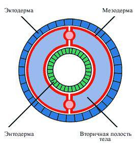
The mesoderm is the middle germ layer, which is a layer of cells and is formed during embryogenesis in multicellular animals (with the exception of sponges, coelenterates).
The mesoderm is located between the primary layers, i.e. ectoderm and endoderm, respectively.

Sources of mesoderm formation
The primary source of mesoderm formation in different representatives of animal species will differ.
- In the vast majority of invertebrates, the middle layer is formed from specialized cells - teloblasts, located in the posterior third of the body of the embryo.
- Part deuterostomes animals, of which fish and amphibians are representatives - certain segments of the wall of the primary intestine serve as the basis of the future mesoderm.
- The other part deuterostomes, for example, birds, reptiles and mammals (to which humans belong), the primary rudiment of the future mesoderm is part of the ectoderm, and after a while it separates into an "independent" leaf.

Development and division of zones
The mesoderm is divided into 4 conditional zones:
- Dorsal zone ... In the course of its development, the mesoderm gradually thickens around the notochord. From these paired thickenings, somites are subsequently formed - dorsal (primary) segments that make up the dorsal part of the mesoderm. Every day, a normally developing human embryo should form two or three pairs of new somites. Thus, after thirty days there are usually 30 pairs of somites. However, do not forget about anatomical features development of each organism, which is characterized by its own minor fluctuations. Despite this, the number of somites will undoubtedly be a certain indicator of the development of the organism.
- Ventral (lateral) zone : the layers of the lateral mesoderm extend on both sides of its dorsal part.
- Intermediate zone of the mesoderm will be located between the previous two and is represented by a narrow connecting zone. V cranial the end of the body of the embryo, this part is involved in the formation of the "temporary" urinary system, and in caudal- participates in the development of a permanent kidney - metanephros.
- Nephrogonadotome - a section of the mesoderm that provides interaction splanchnotome and somites among themselves.
A clear division of the mesoderm into the described parts is characteristic only of its middle third. At its cranial and caudal ends, the mesoderm is represented by poorly differentiated cells that actively move to different departments. Such clusters of cells are called mesenchyme.

Derivatives of mesoderm
- The cells that make up the somite rapidly grow in volume and assume a radial arrangement. Eventually, a cavity appears in the center of the somites - the myocoel, which increases in volume and eventually divides the layer of cells, giving the somite the appearance of a genital ball with powerful walls. At this time, three areas begin to be distinguished inside the somites, the cells of which give rise to the systems of organs and tissues.
- The postero-medial part of the somites is represented by cells from which skeletal muscles will develop. Therefore, it is named myotome.
- The anterolateral part of the somites includes cells that form the connective tissue basis of the skin. Therefore, the name of this part is dermatome, or skin plate.
- The third part of the somite is represented by the so-called sclerotome, the cells of which begin to unite around the neural tube and notochord, eventually forming osteoarticular system.
- Nephrogonadotome differentiates after somites. Ultimately, the cells of this part of the mesoderm give rise to renal tubules nephron, urinary and reproductive ducts.
- Splanchnotomes are divided into two sheets.
- The visceral leaf is adjacent to the endoderm and forms a smooth muscle layer intestines, participates in the formation of blood vessels and blood cells, gives rise to the myocardium and epicardium of the heart, the adrenal cortex.
- The parietal leaf covers the whole from the inside. In the epithelium of the splanchnotomes themselves, the genital ridges are distinguished, which are the future sex glands. Both sheets of the splanchnotome are further involved in the formation of all serous membranes human - peritoneum, pleura, pericardium.
- Part of the mesenchymal cells is also formed from the splanchnotome. These cells give rise to the connective tissue and smooth muscle lining of the internal organs.

Mesoderm(Greek, mesos medium + derma skin) - medium germ layer, or layer; a set of embryonic cells located in the primary body cavity (blastocele) between the outer and inner germ layers (Fig. 1).
M. appears in phylogeny in lower worms and corresponds, according to I. I. Mechnikov, to the peripheral phagocytoblast of coelenterates. Methods for laying and further development of M. are different in different groups animals. In higher vertebrates, M. develops during the second phase of gastrulation, since the future mesodermal material remains after the isolation of the lower (internal) germ layer (in the first phase of gastrulation) as part of the outer layer of the blastodiscus. As a result of cell multiplication and their movement, the mesodermal material forms a longitudinal thickening in the outer layer of the blastodisc in birds and mammals, called the primary stripe (the edges are gradually lengthening in the direction from the caudal to the head region). The anterior, most thickened part of the primary stripe is called the primary, or Hensen's, nodule.

In vertebrates, including humans, M., expanding further and further on both sides of the place of its origin, is subdivided into areas giving rise to different bodies and fabrics. The dorsal part of M., lying on both sides of the notochord and the neural plate (later the neural tube), is metamerically dissected, or segmented, into somites (see), or dorsal segments (Fig. 2). Segmentation begins at the head end and gradually spreads towards the tail end; the number of pairs of segments with the age of the embryo increases (see Metamerism). Somites remain for some time associated with non-segmented ventral M. departments, splanchnotomas, or lateral plates, by means of segmented narrowed intermediate sections - segmental legs, or nephrotomes.
Then the somites differentiate into the medioventral area - the sclerotome, the dorsolateral area - the dermatome, or the skin plate, and the myotome, or the muscle plate, located between them. The relative size of the sclerotome in phylogeny greatly increases; in higher vertebrates this is associated with a more significant development of skeletal tissues.
In the splanchnotomes in the embryos of vertebrates, a slit-like cavity appears - the splanchnocele, in general, or secondary cavity the body, dividing them into two sheets: the outer one is the somatopleura, or the parietal leaf adjacent to the ectoderm, and the inner one is the splanchnopleura, or the visceral leaf adjacent to the endoderm. The mesodermal segments and the notochord form together with the nerve rudiment - the neural plate (more late dates development - a neural tube) so-called. dorsal, or axial, primordium complex, which is characteristic feature all type of chordates. The two anterior segments are separated later than the third and, moreover, in the caudocranial sequence (larval mesoderm). They are represented only by somites, due to which the oculomotor muscles develop. Larval, or larval, segments (I and II) are inherited from the distant ancestors of vertebrates that had the stage of a three-segment larva and underwent metamorphosis. Segmentation of the rest (postlarval) M. gradually spreads to the tail section.
Sclerotomes and dermatomes loosen, forming star-shaped reticular cell junctions and single free cells. Their combination is called entomesenchyme, or mesenchyme (see). The cells that are evicted from the parietal and visceral sheets of splanchnotomes also join the mesenchyme. Parts of splanchnotomes that retain a closed structure after isolation from their mesenchyme are designated as embryonic coelomic epithelium. Myotomes are the main source of skeletal (striated) muscle development.
The mesenchyme, formed from different parts of M., gives rise to all varieties of connective tissue, cartilage and bone formations, hematopoietic organs, blood, lymph, and smooth muscles of the vessels and viscera.
From the nephrotomes, the epithelium of the renal tubules develops. From the splanchnotome, after the cells of the mesenchyme are evicted from it, a large number of various derivatives: mesothelial cover of serous membranes, cardiac muscle. in humans, the adrenal cortex, epithelial parts of the testes and ovaries, unilamellar epithelium uterus and oviducts (in humans, fallopian tubes).
The peripheral parts of the splanchnotomes grow beyond the embryonic scutellum and are part of the embryonic membranes. These parts of the middle germ layer are called extraembryonic M.
In humans and apes, due to the early and intensive development of provisional (temporary) organs, the appearance of extraembryonic M. is shifted to very early stages of uterine life and is strongly altered in comparison with other mammals. Extraembryonic M. is laid in humans earlier than embryonic and independently of it. It fills the blastocyst cavity and underlies its wall, consisting of trophoblast (see), as well as the walls of the amnion, yolk sac and allantois, forming their connective tissue base (see Embryo). The germinal M. developing from the primary stripe is connected with the extraembryonic by its peripheral parts. Therefore, after their reunification (in humans, by the end of the 4th week of embryogenesis), extraembryonic M. appears to be a direct peripheral continuation of splanchnotomes. From extraembryonic M. the first foci of hematopoiesis develop, a kind of connective tissue of a number of provisional embryonic organs, for example, Varton's jelly (see) of the umbilical cord and the lining of the extraembryonic coelom. Disturbances of M.'s differentiation during the period of intrauterine development lead to various malformations (see).
Bibliography: Ivanova-Kazas OM Comparative embryology of invertebrates, vol. 1, p. 54, Novosibirsk- M., 1975; To N about r r e A. G. A short sketch of human embryology, L., 1967; about N e, Embryonic histogenesis, p. 340, L., 1971: S i e w i n g R. Lehrbuch der ver-gleichenden Entwicklungsgeschichte der Tie-re, Hamburg, 1969.
(cm.). From the mesoderm, embryonic rudiments are formed, which serve as a source for the development of muscles, serous cavities, and organs of the genitourinary system.
Mesoderm (from the Greek mesos - middle and derma - skin, layer; synonym: middle germ layer, mesoblast) - one of the three germ layers of multicellular animals and humans on early stages development.
Topographically, the mesoderm occupies an intermediate position between the outer germ layer - ectoderm (see) and the inner - endoderm (see). In the embryos of sponges and most coelenterates, the mesoderm is not formed; these animals remain two-leaved for life. In representatives of higher types of animals, as a rule, the mesoderm appears during the development of the embryo (see) later than the ecto- and endoderm, moreover, it occurs in different animals due to one of these sheets or due to both (respectively, ecto- and entomesoderm are distinguished). In vertebrates, the mesoderm forms as an independent (third) layer of the embryo already in the second phase of gastrulation (Fig. 1).
Rice. 1. Cross section of a vertebrate embryo at the end of the second phase of gastrulation (three germ layers and an axial complex of primordia): 1 - ectoderm (I - cutaneous ectoderm, 2 - neural plate); II - mesoderm (3 - mesoderm, 4 - chordal cord); III - endoderm.
In a series of vertebrates, there is a gradual change in the way the mesoderm is formed. For example, in fish and amphibians, it occurs in the area bordering between the ento- and ectoderm, formed by the lateral lips of the primary mouth (blastopore). In birds, mammals, and humans, the cellular material of the future mesoderm is first collected in the form of a primary strip as part of the outer germ layer (in humans, on the 15th day of intrauterine development), and then plunges into the gap between the outer and inner layers and lies on both sides of the primordium of the dorsal string (chord), entering with it and the primordium nervous system into the axial complex of primordia. The parts of the mesoderm (axial) closest to the notochord primordium are part of the body of the embryo and take part in the formation of its permanent organs. The peripheral areas grow in the interval between the marginal parts of the ecto- and endoderm and are part of the auxiliary temporary organs of the embryo - the yolk sac, amnion and chorion.
The mesoderm of the trunk of the embryo of vertebrates and humans is divided into dorsal sections - dorsal segments (somites), intermediate - segmental legs (nephrotomas) and ventral - lateral plates (splanchnotomes). Somites and nephrotomas are gradually segmented from front to back (in humans, the first pair of somites occurs on the 20-21st day of intrauterine development, the last, 43 or 44th, pair - by the end of the 5th week). Splanchnotomas remain unsegmented, but are split into parietal (parietal) and visceral (visceral) sheets, between which there is a secondary body cavity (whole). Somites are subdivided into dorsolateral regions (dermatomes), medioventral (sclerotomes), and intermediate between them (myotomes). Dermatomes and sclerotomes, acquiring a looser arrangement of cells, form a mesenchyme (see). Many cells of the mesenchyme are also evicted from splanchnotomes. A diagram of organogenesis in the embryo of a higher vertebrate is shown in Fig. 2. So, in particular, from myotomes, an arbitrary striated muscle skeletal muscles. Nephrotomas give rise to the epithelium of the kidneys, oviducts and uterus. Splanchnotomas turn into a single-layer squamous epithelium lining the whole - the mesothelium (see). They also form the adrenal cortex, the follicular epithelium of the gonads and the muscle tissue of the heart.

Rice. 2. Scheme of organogenesis in the embryo of a higher vertebrate (the names of tissue derivatives are put in brackets after the name of the corresponding anlage): 1 - cutaneous endoderm (epidermis); 2 - ganglion plate (sensitive and sympathetic neurons, peripheral neuroglia, chromatophores); 3 - neural tube (neurons, neuroglia); 4 - chord; 5 - dermatome (connective tissue basis of the skin); 6 - myotome (musculoskeletal tissue); 7 - sclerotome (cartilaginous and bone tissue); 8 - nephrotome (renal epithelium); 9 - parietal sheet of the splanchnotome (mesothelium); 10 - visceral sheet of the splanchnotome (mesothelium, heart muscle tissue); 11 - intestinal endoderm (intestinal epithelium); 12 - mesenchyme (connective tissue, blood, smooth muscle tissue); 13- extraembryonic ectoderm (amnion epithelium); - 14 - aortic endothelium; 15 - yolk endoderm (yolk sac epithelium); 16 - in general.
See also Germ leaflets.
Embryogenesis - difficult process, which is characterized by the gradual formation of organs and tissues. In most multicellular organisms, the embryonic rudiment consists of three layers: ectoderm, endoderm, mesoderm. What is mesoderm? Both the chitinous skeleton of arthropods, and the epidermis of the skin, and the nervous system are of ectodermal origin. Digestive, endocrine and respiratory system are formed from endoderm. What organs and tissues does the mesoderm give rise to? How is it formed?
What is mesoderm. Definition
Any tissue or organ system is formed from a specific layer of cells in the embryo. What is mesoderm? In biology, the definition sounds like this: it is one of the germ layers, from which a number of organs and tissues are formed in the process of embryogenesis. The second name of the mesoderm is mesoblast. The formation of this layer is typical for most multicellular animals (exception: the Sponge type and the Intestinal type).

The mesoderm is located between the ectoderm and endoderm. Each of the nearby germ layers can take part in the formation of the mesoblast. Accordingly, by origin, two types of median germ layer are distinguished: entomesoderm, exomesoderm. There are also situations when both structures take part in the formation of the mesoblast at once.
The mesoderm as an independent one is formed at the stage of gastrulation.
The formation of the mesoderm. Formation features
What is mesoderm? In biology, it is generally accepted that each organ of a multicellular animal in embryogenesis is formed by one of the germ layers. The formation of the mesoderm is a characteristic aramorphosis, since for the first time a true median embryonic layer is formed in them. Type Sponges and are representatives of two-layer animals: in embryogenesis, only ectoderm and endoderm are formed.
How does the mesoderm form?
There are three ways of mesoblast formation.

Mesoderm structure
What is mesoderm? This is not just an accumulation of identical cells, but a germ layer differentiated into several functional divisions. The division of the mesoblast occurs gradually, as a result of which the following areas are distinguished:
- Somites are paired ribbon-like formations, between which a whole is formed - a secondary body cavity. They are also preserved in arthropods.
- The notochord bud is a section of the mesoderm that develops into a notochord in the future. Distinctive feature vertebrates.
- In vertebrates, a sclerotome, dermatome, and myotome are formed from each somite.
- Splanchnotomas - lateral plates, which are dissected into two separate layers: inner and outer. Between them, a whole is formed in vertebrates.
- Nephrotomes are paired structures that connect splanchnostomes.
Having studied each part of the germ layer, scientists were able to determine what the mesoderm is and understand what functions it performs.
Histogenesis
The mesoderm gives rise to several types of tissue.
- The parenchyma of flatworms, which fills the space between the organs. Formed from the mesoderm.
- Some epithelial tissue covering organs outside. This includes secretory cells, endocrine and exocrine glands.
- Loose fibrous and dense fibrous connective tissues are formed from the mesoderm. Including collagen and elastic fibers are formed.
- also formed from the mesoderm.
- Bone and cartilage tissues, their constituent elements are of mesodermal origin.
- By analogy with the blood cells, the mesoderm also takes part in the formation of cells of the immune system.
- All types of muscle tissue. Smooth muscles are embedded in the walls of most organs. Cross-striped fibers are structural elements skeletal muscles. Do not forget about the striated heart muscle tissue, which forms the musculature of the heart.
Organogenesis
The tissues form organs, so it is not difficult to guess which ones are of mesodermal origin. The classification is given by mesoderm sites:
- dermatomes - form the dermis of the skin (the skin contains sweat and sebaceous glands);
- the passive part of the musculoskeletal apparatus (skeleton) is formed from the sclerotomes;
- from the myotome, respectively, - the active part of the musculoskeletal system (muscles);
- splanchnostomas give rise to mesothelium - a single-layer epithelium that lines the secondary body cavity;
- nephrostomy cells form the excretory and reproductive systems.

mesodermal origin
It is worth mentioning those organs that are lost at different stages of ontogenesis after the fulfillment of their functions. They are called provisional. These include:
- Amnion is one of the membranes of the embryo that performs several vital functions at once. The first is to create an aquatic environment for the development of the fetus. This is explained by the fact that the formation of an organism must take place in water. For vertebrates living on land, water in this case is a limiting factor, therefore, this shell was formed in the process of evolution. Also Amnion protects the fetus from mechanical damage, maintains a constant environment by maintaining the concentration of salts at a constant level, and also protects the embryo from the effects of toxic substances.
- Allantois is another organ of the embryo that performs the functions of nutrition and respiration at the same time. By origin, it is an outgrowth of the yolk sac, which means that it is also formed by the cells of the endoderm and mesoderm. In humans, allantois is less developed than in other vertebrates, but they pass through it blood vessels, which then fall into the tissue of the umbilical cord.
- Yolk sac. This temporary organ contains the nutrients needed for the development of the fetus. Cells of both mesoderm and endoderm are involved in the formation of the yolk sac. An interesting feature of the organ is the formation of the very first blood cells of the body in it.
- Umbilical cord (umbilical cord) - connects the embryo and the placenta.
- Chorion - the shell of the embryo, with the help of which attachment to the uterus and the formation of the placenta occurs.
- The placenta is the only organ in humans tissue-formed two organisms: mother and fetus. From the mother's blood, the embryo receives nutrients and oxygen through the placenta.
Functions of the mesoderm
We examined what the mesoderm is. What are the functions of this germ layer?
The development of the mesoderm allowed the flatworms to fill the gaps between the organs with parenchymal tissue. More perfect organisms do not have parenchyma, but the principle is similar: tissues of mesodermal origin form boundary layers between organs. The most important function of the mesoblast is the formation of temporary organs in the embryo (allantois, umbilical cord, placenta, etc.). Also, mesoderm cells form the tissues of the internal environment: blood and lymph.

Conclusion
Now in to the fullest you can explain what the mesoderm is. Its formation allowed the animals to move to a new stage of development, as evidenced by the origin of many organs and tissues. In addition, the formation of the amniotic membrane caused a qualitative leap in the development of vertebrates. Hence, the mesoderm is an important evolutionary element.
mesoderm
The mesoderm (synonym mesoblast) is the middle germ layer, consisting of cells that lie in the primary body cavity between the ectoderm (see) and endoderm (see). From the mesoderm, embryonic rudiments are formed, which serve as a source for the development of muscles, epithelium of serous cavities, and organs of the genitourinary system.
See also Fetus.
The mesoderm (from the Greek mesos - middle and derma - skin, layer; synonym: middle germ layer, mesoblast) is one of the three germ layers of multicellular animals and humans in the early stages of development.
Topographically, the mesoderm occupies an intermediate position between the outer germ layer - ectoderm (see) and the inner - endoderm (see). In the embryos of sponges and most coelenterates, the mesoderm is not formed; these animals remain two-leaved for life. In representatives of higher types of animals, as a rule, the mesoderm appears in the process of development of the embryo (see) later than the ecto and endoderm, moreover, it occurs in different animals due to one of these leaves or due to both (respectively, ecto and entomesoderm are distinguished). In vertebrates, the mesoderm forms as an independent (third) layer of the embryo already in the second phase of gastrulation (Fig. 1).
In a series of vertebrates, there is a gradual change in the way the mesoderm is formed. For example, in fish and amphibians, it occurs in the area bordering between the ento and ectoderm, formed by the lateral lips of the primary mouth (blastopore). In birds, mammals, and humans, the cellular material of the future mesoderm is first collected in the form of a primary strip as part of the outer germ layer (in humans, on the 15th day of intrauterine development), and then plunges into the gap between the outer and inner layers and lies on both sides of the primordium of the dorsal string (chord), entering with it and the primordium of the nervous system into the axial complex of the primordia. The chord (axial) parts closest to the rudiment of the chord are part of the body of the embryo and take part in the formation of its permanent organs. The peripheral areas grow in the interval between the marginal parts of the ecto and endoderm and are part of the auxiliary temporary organs of the embryo - the yolk sac, amnion and chorion.
The mesoderm of the trunk of the embryo of vertebrates and humans is divided into dorsal sections - dorsal segments (somites), intermediate - segmental legs (nephrotomas) and ventral - lateral plates (splanchnotomes). Somites and nephrotomas are gradually segmented from front to back (in humans, the first pair of somites occurs on the 20-21st day of intrauterine development, the last, 43 or 44th, pair - by the end of the 5th week). Splanchnotomas remain unsegmented, but are split into parietal (parietal) and visceral (visceral) sheets, between which there is a secondary body cavity (whole). Somites are subdivided into dorsolateral regions (dermatomes), medioventral (sclerotomes), and intermediate between them (myotomes). Dermatomes and sclerotomes, acquiring a looser arrangement of cells, form a mesenchyme (see). Many cells of the mesenchyme are also evicted from splanchnotomes. A diagram of organogenesis in the embryo of a higher vertebrate is shown in Fig. 2. So, in particular, from myotomes, an arbitrary striated muscle tissue of skeletal muscles develops. Nephrotomas give rise to the epithelium of the kidneys, oviducts and uterus. Splanchnotomas turn into a single-layer squamous epithelium lining the whole - the mesothelium (see). They also form the adrenal cortex, the follicular epithelium of the gonads and the muscle tissue of the heart.
Rice. 2. Scheme of organogenesis in the embryo of a higher vertebrate (the names of tissue derivatives are put in brackets after the name of the corresponding anlage): 1 - cutaneous endoderm (epidermis); 2 - ganglion plate (sensitive and sympathetic neurons, peripheral neuroglia, chromatophores); 3 - neural tube (neurons, neuroglia); 4 - chord; 5 - dermatome (connective tissue basis of the skin); 6 - myotome (musculoskeletal tissue); 7 - sclerotome (cartilage and bone tissue); 8 - nephrotome (renal epithelium); 9 - parietal sheet of the splanchnotome (mesothelium); 10 - visceral sheet of the splanchnotome (mesothelium, heart muscle tissue); 11 - intestinal endoderm (intestinal epithelium); 12 -
C1 task on the exam in chemistry
Preparing for the exam in German
The structure of the examination paper consists of two blocks
What is the exam in Russian
What is VLOOKUP and why should they be done?