Each peripheral nerve consists of a large number of nerve fibers united by connective tissue sheaths (Fig. 265- A). In the nerve fiber, regardless of its nature and functional purpose, they distinguish axial cylinder- cylindroaxis, covered with its own sheath - axolemma and the nerve sheath - neurolemma. If the latter contains a fat-like substance - myelin, the nerve fiber is called pulp or myelin neurofibra myelinate, and if it is "absent," fleshy or amielin- neurofibra amyelinata (bare nerve fibers - neurofibria nuda).
The value of the pulp is that it promotes better conduction of nervous excitement. In the non-fleshy nerve fibers, excitation is carried out at a speed 0,5-2 m / s, while in pulp fibers - 60-120 m / s. "By diameter, individual nerve fibers are subdivided into thick pulpy fibers (from 16-26 μm in horses, ruminants up to 10-22" μm in a dog) "- efferent somatic; medium pulpy (from 8-15 μm in horses, ruminants up to 6-8 μm in a dog) - afferent somatic; thin (4--8 μm) ^ -y efferent vegetative (Fig. 265- B).
Non-fleshy nerve fibers are part of both somatic and visceral nerves, but in quantitative terms there are more of them in the autonomic nerves. They differ both in diameter and in the shape of the neurrolem nuclei: 1) low-fleshy, or non-fleshy, fibers with a rounded core (fiber diameter 4-2,5 μm, core size 8X4 ", 6μm, distance between nuclei 226-345 μm); 2) low-fleshy or non-fleshy fibers with an oval-elongated shape of the neurolemma nuclei (fiber diameter 1-2,5 μm, core size 12,8 X 4 μm, distance between nuclei 85- 180 μm); 3) fleshy fibers with a fusiform shape of the nuclei of the neurolemma (fiber diameter 0,5-1,5 μm, core size 12,8 NS 1,2 μm, ras-
A .56
Rice. 265. The structure of the peripheral nerve? A - nerve on a transverse section: / - epineurium; 2 - perineurium; 3 - endoneurium! 4 - neurofibra myelinata; 5 - cylindraxis; 5 - the composition of the nerve fibers in the somatic nerve of the sheep; 1, 2, 3 - neurofibra myelinata; 4 - neurofibra amyelinata; 5i6, 7 - neurofibra nuda; a - temmocytus; e- incisio myelini; with - isthmus nodi.
standing between fibers 60-120 microns). In animals of different species, these indicators may be different.
Sheaths of the nerve. Nerve fibers extending from the brain, by means of connective tissue, are combined into bundles that form the basis of peripheral nerves. In each nerve, connective tissue elements participate in the formation: a) inside the bundle base - endoneurium, located in the form of loose connective tissue between individual nerve fibers; b) the connective tissue sheath covering individual groups of nerve fibers, or perineurium- perineurium. In this shell, a double layer of flat epithelial cells of "ependymal nature is distinguished from the outside, which form a perine-oral vagina around the nerve bundle, or perineural space- spatium peri-neurii. From the basilar inner layer of the lining of the perineural vagina into the depths of the nerve bundle, connective tissue fibers depart, forming bundle fibers inside perineural septa- septum peri-neurii; the latter serve as a place! passage of blood vessels, as well as participate in the formation of the endoneurium. ^
The perineural sheaths accompany bundles of nerve fibers along their entire length and divide as the nerve divides into smaller branches. The cavity of the perineural vagina is reported by the subarachnoid and subdural spaces of the spinal cord or brain and. contains a small amount of cerebrospinal fluid (a neurogenic route of penetration of the rabies virus into the central parts of the nervous system).
Groups of primary nerve bundles by means of dense loose connective tissue are combined into larger secondary and tertiary bundles of nerve trunks and constitute the outer connective tissue sheath in them, or epineurium- epineurium. In the epineurium, in comparison with the endoneurium, there are larger blood and lymphatic vessels - vasa nervorum. Around the nerve trunks there is a certain amount (depending on the place of passage) of loose connective tissue, which forms an additional near-nerve (protective) sheath along the periphery of the nerve trunk - paraneural T. In the immediate vicinity of the nerve bundles, it is transformed into an epineural sheath.
16-341 449
REGULARITIES OF STROKE AND BRANCHING OF NERVES
The topography and branching of peripheral nerves has much in common with topo graphics and branching of blood vessels, with which they often pass together, forming neurovascular bundles. Their joint passage is due to the peculiarities of the development of the organs for which they are intended, the area of distribution and the conditions of functioning. Being located in one common connective tissue case, the blood vessels ensure the creation of an optimal temperature regime for conduction of nerve impulses, as well as for the nutrition of nerve trunks. In addition, some other features are characteristic of the peripheral nerves.
]. "The spinal nerves from the spinal cord depart metamerically in accordance with the division of the bone base and are subdivided into cervical, thoracic, lumbar, sacral and caudal. Cranial nerves depart from the medulla oblongata (from XII on V pair) and midbrain (IV and III pairs). I and II Cranial pairs of nerves occupy a special position in this respect, being the nerve tracts of the most important sense organs.
2. Each spinal nerve has two root - dorsal and ventral- radix dorsalis et ventralis. On the dorsal root is spinal ganglion- ganglion spinale. Both roots at the exit from the spinal canal are connected to a common nerve barrel- spinal nerve - item spinalis, "containing sensory, motor and sympathetic fibers. The cranial nerves depart mainly with one root, corresponding to the dorsal or ventral root of the spinal nerve.
3: All efferent (motor) nerve fibers exit from the ventral columns of the gray medulla of the spinal cord and from the "corresponding motor nuclei of the medulla oblongata and midbrain (III, IV, VI, XI, XII). On the spinal cord, they form ventral motor roots.
All afferent (sensory) nerve fibers consist of neurites of the cells of the spinal nodes and, accordingly, the ganglia of the cranial nerves (pairs V, VII, VIII, IX and X). Consequently, all the bodies of receptor (sensory) neurons lie outside the spinal cord and brain.
Each spinal nerve, upon exiting the spinal canal, gives off a white connecting branch - ramus (g) communicans albus - into the sympathetic trunk, "a branch into the membranes of the spinal cord" - g. meningeus, then receives a gray connecting branch - g. communicans griseus - from the sympathetic trunk and is divided into dorsal and ventral branches - dorsalis et ventralis - according to the differentiation of the trunk muscles into dorsal and ventral muscle cords with their vessels. Each of these branches, in turn, is divided into medial and lateral branches - rnedialis et lateralis - for the muscles and skin, which is also due to the division of muscle cords into lateral and medial layers.
together with the corresponding part of the spinal cord forms a nerve segment - neurotic- neurotom. Neurotomas are more pronounced where there is a clear segmentation in the skeleton and muscles, for example, in the thoracic region of the trunk.
6. When the myotomes are displaced in the process of evolution, the branches of the corresponding neurotomes innervating them are displaced after them. So, the diaphragmatic nerve - p. Phrenicus, originating from the 5-7th cervical neurotomy, approaches the diaphragm through the entire chest cavity; or, for example, the accessory nerve - item accessorius - leaves the spinal canal through a torn opening in the skull, and goes to the cervical region to innervate the brachiocephalic, trapezius and sterno-maxillary muscles.
In the area of nerve discharge in the limb, the brachial and
Fig. 266. Zones of distribution of cutaneous nerves: / - infraorbital N; G - subblock N.; 2 - frontal N.; 2 1
1 - zygomatic N .; 3 - dorsal branches of the cervical N.; 4 - dorsal branches of the chest N.; 5 - ilio-hypogastric N .; 6 - iliac groin N.; 7 - cranial cutaneous gluteal N; 8 - middle cutaneous gluteal n.; - 9 - tail n.; 10 - perineal N.; 11 - Caudal cutaneous gluteal N; 12 - tibial N.; 13 - plantar cutaneous nerves of the foot; 14 - low-boric superficial n.; 15 - cutaneous lateral nerve of the leg; 16 - cutaneous lateral nerve of the thigh; 17 - external shameful N .; 18 - n. safenus; 19 - cutaneous medial n. feet; 20 - ventral branches of thoracic N; 21 - elbow N.;, 22 - middle n .; 23 - musculocutaneous N .; 24 - radial surface n,; 25 - axillary N.; 26 - ventral cervical N.; 27 -
mandibular n.
lumbosacral nerve plexuses - plexus brachialis et lumbosacral, and from them nerves already originate, heading to certain muscle groups. Typically, both the nerves and muscles of the limbs are multi-segment. Nerve plexuses are also found in the neck, which is also explained by the complex origin of the cervical muscles. Connecting branches between individual nerves -rr. communicantes - indicate the origin of individual nerves from several neurotomes.
7. The sensory nerves, although mainly correspond to the skin segments - dermatomes, innervate not only the area of their segment, but also enter the adjacent dermatomes. Therefore, anesthesia of any skin segment. (dermatome) is possible only when three adjacent neurotomes are turned off (Fig. 266).
SPINAL NERVES
Spinal nerves "- nervi spinales - are divided into cervical (C), thoracic (Th), lumbar (L), sacral (S) and caudal (Co) (according to the division of the spinal column).
NECK NERVES
Cervical nerves - nn. cervicales - in the amount of 8 pairs exit through the intervertebral foramen [the first pair (C I) comes out through the intervertebral foramen of the atlas, the second pair (C II) through the intervertebral foramen behind the atlas, and the eighth pair (C VIII) - behind the 7th cervical vertebra ]. Every cervical nerve receives gray branch- griseus, including C VIII-VII - from the stellate node, C VI-III (II) -j-ot of the vertebral nerve and C I (II) - from the cranial cervical sympathetic node. Having received a gray branch and given sheathing branch- Mr. meningeus, the spinal nerve is divided into dorsal and ventral branches- rr. dorsales et ventrales. The dorsal medial branches go along the medial surface of the podostasis "muscles of the head and neck, and the lateral ones - along the medial surface of the neck muscles - plaster-like and longest. The dorsal medial branch of the first cervical nerve is called the greater occipital nerve - item occipitalis major, which branches into short muscles of the occipital and axial joints, as well as in the skin of the occipital region and the caudal muscles of the ear shell.
Individual ventral branches of the cervical nerves are characterized by a special course and, accordingly, receive special names. The ventral branch of the first "cervical nerve connects with the hypoglossal and ventral branch of the second cervical nerve, forks in the muscles of the neck. The ventral branch of the second cervical nerve has connections with C I, C III, accessory nerve. From it beret Start large ear nerve - n. auricularis magnus, which branches out in the skin of the base of the head, muscles * of the auricle and here It has connections with branches of n. facialis. The continuation of the ventral branch of C II is transverse nerve of the neck- item transversus colli; having received a connecting branch from C III, it branches in the skin, having connections with the skin branches of the neck - NS. facialis.
The phrenic nerve - n. phrenicus - comes from C (V), VI and VII. Medially "from the scalene muscle and the subclavian artery, it goes into the chest cavity and branches out in the diaphragm.
Supraclavicular nerve- item supraclavicular is - comes from C VI, forks in the skin of the shoulder joint, shoulder and dewlap. The ventral branches of the last 3 (4): the cervical nerves take part in the formation of the brachial plexus, from which the nerves for the shoulder girdle and the free part of the thoracic limb come out.
SHOULDER! PLEXUS
Brachial plexus - plexus brachialis - formed by two trunks - trunci plexus - from the ventral branches C VI, VII and C VIII, Th I (II). It lies ventrally from the "scalene muscle and medially from the scapula. Nerves that innervate the region of the shoulder girdle, the muscles of the scapula come out of it. and free section of the limb (Fig. 267).
Dorsal nerve scapula-p. dorsalis scapulae (15) -double, -departing from С V and Vi. Both nerves go to the rhomboid muscle - one along the medial surface, and the other in the thickness of the cervical part of the ventral dentate muscles, to which they send branches. It has connecting branches with a long pectoral nerve.
Long pectoral nerve- n. thoracicus longus - originates in two branches from C VII-VIII, which, having united, are directed caudally and branch into the ventral dentate Muscle.
Suprascapular nerve- n. suprascapularis (1) - formed from C VI and VIi , iHfleT together with the suprascapular artery into the abdominal and extraspinal muscles and into the scapula.
Subscapular nerves- nn. subscapulars (b) - in the amount of 2-4
start from C VI and go to the muscle of the same name, giving off branches to the large round muscle and the periosteum of the medial surface of the scapula. ... I ..
The thoracic nerve - n. Thoracodorsal (7) - originates along with the subscapularis or axillary nerves from C VI ^ -VII (in ungulates C VII-VIII) and goes to the latissimus dorsi muscle, giving off branches to the large circular muscle along its course.
Axillary nerve - n. Axillaris (4) - starts from C VII-VIII, "together with the brachial circumferential lateral artery penetrates between the subscapularis and large round muscles inward and, giving away the muscle branches v small round and deltoid muscles (in dogs and horses also in the capsular), extends to the lateral surface of the shoulder. Here, the cranial lateral cutaneous nerve of the shoulder departs from it - item cutaneus brachii lateralis cranialis - and continues to the forearm, where it is called the cranial cutaneous nerve of the forearm - item cutaneus antebrachii cranialis, here it branches, reaching the wrist (V.I. ).
Radial nerve - n. Radialis (10) - the largest nerve of the extensor surface of the thoracic limb. It begins with nerve bundles from C VII - C VIII and Th I, passes between the heads of the triceps muscle of the shoulder, where it gives them muscle branches. Bending around the humerus from the caudal surface in the laterodistal direction, the radial nerve v the area of the elbow joint gives caudal lateral cutaneous nerve of the shoulder -
h. cutaneus brachii lateralis caudalis - and is divided into superficial and deep branches. Deep branch- Mr. profundus - is divided into muscle branches that branch out in the extensors of the forearm. Surface branch - g. superficialis (Fig. 268- 10), giving away the lateral cutaneous nerve of the forearm -NS. cutaneus antebrachii lateralis, and in carnivores and pigs also lateral and medial branches, continues distally and in the wrist is divided into common dorsal digital nerves- nn. digitales dorsales commu-

Rice. 268. Hand nerves: A - dogs; B - pigs; V - cows (from the dorsal surface); G - horses; D - dogs; E - pigs; F - cows (from the palmar surface); 3 - item muscu-locutaneus; 5 - n. Medianus; 10 - g. superficialis n. radialis; 11 - item ulnaris; 11" - dorsalis n. ulnaris; 13 - item digitales palmares communes; 13" - r. communicans; 14- n. digitalis palmaris proprius (in a horse - lateralis); 15 - nn. digitales dorsales proprii; 16 - nn. digitales dorsales communes; / - V - fingers.
hes (I-IV - in carnivores, II-IV - in pigs, II-III - in ruminants; horses do not have them), which continue into the dorsal digital nerves proper. In carnivores, along with the common dorsal digital nerves, it departs from the superficial branch first non-axial dorsal digital nerve- n. digitalis dorsalis I abaxialis.
Musculocutaneous nerve- n. musculocutaneus (3) - originates from C VI-VII and, having given up the proximal branch - r. proximalis - into the coracoid-brachial and biceps muscles, together with the median nerve in ungulates forms axillary loop- ansa axillaris.
In carnivores, the musculocutaneous nerve passes along the medial surface of the shoulder along the biceps muscle (in ungulates, it passes together with the focal nerve from the axillary loop to the distal third of the forearm, where it regains its independence). Having given the distal muscular branch to the brachial muscle and exchanging the connecting branches of the focal nerve (in carnivores), the musculocutaneous nerve continues as a medial cutaneous nerve of the forearm- n. cutaneus? antebrachii me-dialis. "
Median nerve- item medianus (5) - originates from C VII-VIII, Th I, passes along the medial surface of the shoulder (in ungulates, together with the musculocutaneous nerve) and in the area of the elbow joint gives off muscle branches to the circular pronator and superficial finger flexor ( in carnivores), the flexors of the wrist and the deep digital flexor, in which it has intramuscular connections with the branches of the ulnar nerve. Then, giving the interosseous forearm nerve- item interosseus antebrachii, descends to the distal end of the forearm and is divided into common palmar digital nerves - nn. digitales palmares communes I-III (carnivores), II-III (pig, ruminants), and in a horse on the medial and lateral palmar nerves - nn. .palmares medialis et lateralis, which correspond to the second and third common palmar digital nerves (13). Common palmar digital nerves from the metacarpal bones pass into the corresponding palmar digital nerves proper - n. Digitalis palmaris proprius I-IV (carnivorous), II-IV (pig, ruminant) and in a horse to the lateral and medial palmar digital nerves - nn. digitales palmares lateralis et medialis (14).
Ulnar nerve- n. ulnaris (11) - formed due to C VIII and Th I (in a horse and dog and Th II), passes along the medial surface of the shoulder towards the elbow tubercle, giving off along its course caudal cutaneous nerve of the forearm- item cutaneus antebrachii caudalis, which reaches the palmar surface of the wrist, and muscle branches in the caudal muscles of the forearm. Above the wrist, the ulnar nerve divides into dorsal and palmar branches.
Dorsal branch.- dorsalis "- is divided; into the common dorsal digital nerve - item digitalis dorsalis communis" IV (carnivores, pigs, ruminants) and V non-axial dorsal digital nerve - item digitalis dorsalis V abaxialis (carnivores, pig), which continue distally and de-; lie on their own dorsal digital nerves - nn. digitales dorsales proprii IV-V (cat, pig, ruminants). In the horse, the dorsal ramus forks in the skin of the dorsolateral surface of the wrist and metacarpus. ,
Palmar branch- Mr. palmaris, in turn, is divided on superficial and deep branches.
Surface branch- Mr. superficialis - is divided into two branches. One goes as a common dorsal digital nerve - item digitalis palmaris communis IV (carnivores, pigs, ruminants), and in a horse III, or the lateral digital nerve - item palmaris lateralis, in the formation of which the palmar lateral branch of the median nerve also participates. In the middle of the metacarpal "bone, the lateral palmar nerve in a horse takes a connecting branch from the medial palmar nerve. In the area of the metacarpopalatine joint, the common digital palmar nerve is divided into the digital nerve itself, axial to V (carnivores, pigs," ruminants), non-axial to IV ( carnivores, pigs, ruminants) and the lateral finger in a horse, a dorsal branch departs from it (15) for the laterodorsal surface of the finger. The second branch, extending from, Mr. superficialis, is found in carnivores and pigs and innervates their fifth finger-: item digitalis palmaris V abaxialis.
Deep branch- Mr. profundus, extending from the palmar branch of the ulnar nerve, is divided into palmar metacarpal "nerves - nn. metacarpei palmares (dog, horse), branching into the interosseous and vermiform muscles, reaching the distal end of the metacarpus. In other animals, it is short and branches in the area wrists.
Thoracic cranial nerves- nn. pectorales craniales (2) - 3-4 branches are formed from the medial surface of the brachial plexus from C VI-VIII and are sent to the superficial pectoral muscles, v which and branch out.
Thoracic caudal nerve-t n. pectoralis caudalis (8) - originates from the medial surface of the brachial plexus from C VIII-Th I (in a dog and horse and from Th II), giving away a muscular branch to the caudal superficial pectoral muscle, continues as lateral pectoral nerve,-NS. thoracicus lateralis (8) - for innervation of the side wall of the chest. In a horse, a ventral branch is separated from it, passing along the superficial pectoral muscle in the caudal direction, being lost in the skin of the lateroventral surface of the chest wall.
Content
The central nervous system is the brain and spinal cord, which are responsible for the proper functioning of the body. For this, there is a peripheral nervous system, consisting of nerves, receptors, nodes, sensitive cells that transmit signals from the whole organism to the central NS. Many diseases, from sciatica to vertebral lesions, are specifically associated with lesions of the PNS, which does not have its own defense mechanisms or blood-brain barrier.
What is the Peripheral Nervous System
The structure of the peripheral nervous system includes nerve endings, ganglia (localized bundles of neurons in all parts of the body), sensory organs, nerves, nerve nodes. The PNS itself is conditionally divided into several subsystems, which, in the complex of their actions, transmit information about the world around, the state of the organism to the brain.
In fact, the peripheral nervous system is responsible for interacting with the outside world, transmitting information to the brain, adequate functioning of internal organs, and correct reaction to external stimuli after receiving a response signal from the brain (for example, the release of adrenaline at a moment of danger). Unlike the central nervous system, this part is not protected by anything and is subject to a large number of dangers.
Classification
It is customary to divide the peripheral part of the nervous system into several subsystems, depending on the direction of its action (external or internal world), the place of communication with the central nervous system, and the temporary moment of work. However, they interact so closely that it is often difficult to attribute a process to a separate system. Medical division of parts of the peripheral nervous system according to the main types of functioning:
- Somatic. The system ensures the independent functioning of the body in the surrounding world, movement, muscle control. This also includes the sense organs as a way of perceiving the environment, full-fledged interaction with it.
- Vegetative (visceral). This part of the peripheral nervous system is responsible for internal organs, glands, blood vessels and partially for some muscles.
It is also customary to divide the vegetative system into parts of the brain and spinal cord, the centers of which correspond to the nerve endings, and the periods of functioning:
- sympathetic system: responsible for the pulse, gastric motility, respiration, blood pressure, the work of small bronchi, pupil dilation, etc. served by sympathetic fibers starting in the lateral horns of the spinal cord, activated at the moment of stress;
- parasympathetic system: functionally opposed to the previous one, for example, it is responsible for the constriction of the pupil (most organs receive both signals from both parts of the nervous peripheral system), the signals are received from the centers in the sacral spinal cord and the brain stem, it works at the moment of rest of the person.
Functions
The peripheral nervous system consists of paired nerves of three key groups: cranial, spinal, and peripheral. They are responsible for the transmission of impulses, commands to the body, organs from the brain and its feedback with the outside world. Each group of endings is responsible for specific functions, therefore, their damage leads to the loss of a particular ability or its modification. Here are just some of the vital processes that the PNS controls:
- the production of hormones responsible for psychological reactions (excitement, joy, fear);
- sensory definition of the world (visual perception, tactile sensations, taste, smell);
- responsible for the functioning of the mucous membranes;
- coordination in space (vestibular apparatus);
- is responsible for the functioning of the genitourinary, circulatory system, intestines;
- production of peptides, neuropeptides;
- contraction of tendons;
- responsible for regulating heart rate and many others.

Peripheral nerves
This is a group of mixed functionality bundles. Unlike other elements of the peripheral nervous system, these nerves are formed into powerful channels isolated by connective tissue. Because of this feature, they are much more resistant to damage, but their injury poses great problems for the body systems. Peripheral nerve bundles are divided into three groups according to the place of attachment to the lumbar column:
- shoulder;
- lumbar;
- sacral.
Dorsal nerves of the cervical spine
PNS is paired nerves in the amount of 12 pairs, which are responsible for the transmission of impulses, commands to the body, organs from the brain and feedback from the outside world. Each group of nerve endings is responsible for specific functions, therefore, their damage leads to the loss of a particular ability or its modification. 12 pairs of cerebral (cranial) nerves of the PNS:
- Olfactory.
- Visual (responsible for the pupillary response).
- Oculomotor.
- Block (responsible for controlling eye movement).
- Ternary - transmits signals from the face, controls the chewing process.
- Abductor (takes part in eye movement).
- Facial - controls the movement of facial muscles, is responsible for the perception of taste.
- The vestibular cochlear. Responsible for the transmission of auditory impulses, a sense of balance.
- Glossopharyngeal.
- Wandering - responsible for controlling the muscles of the pharynx, larynx, organs in the chest, peritoneum.
- Dorsal - responsible for the work of the muscles of the neck and shoulders.
- Sublingual.
Brachial plexus
This is a complex of 4-8 cervical and 1-2 spinal nerves, which are responsible for the innervation of the skin of the hands and the functioning of muscles. The plexus itself is localized in two areas: in the axillary fossa and the lateral triangle of the neck. The short and long branches of nerves are made up of channels, each of which is responsible for an individual muscle and nerve perception of the skin, muscles and bones.

Neurotransmitters
It was believed that the exchange of signals between nerve endings, the central nervous system, and the peripheral nervous system occurs through electrical signals. But research has shown that there are not enough of them, and chemicals have been identified - neurotransmitters. Their purpose is to strengthen the connections between neurons and modify them. The number of neurotransmitters has not yet been fully determined. Some of the notable ones are:
- glutamate;
- GABA (gamma-aminobutyric acid);
- adrenalin;
- dopamine;
- norepinephrine;
- serotonin;
- melatonin;
- endorphins.
Diseases of the peripheral nervous system
PNS is so extensive and performs such a number of functions that there are a great many options for its damage. It should be remembered that this system is practically not protected by anything, except for its own structure and surrounding tissues. The central nervous system has its own protective and compensatory mechanisms, and the peripheral nervous system is subject to mechanical, infectious, toxic effects. Diseases of the peripheral nervous system:
- vertebral lesions: reflex syndromes, cervicalgia, cervicocranialgia, cervicobrachialgia, radicular syndromes, radiculitis of the roots, radiculoischemia, thoracalgia, lumbodynia, lumbago, amyotrophy, funiculitis, plexitis;
- lesions, inflammation of the nerve roots, plexuses, nodes: meningoradiculitis, plexitis, plexus trauma, ganglionitis, truncitis;
- multiple lesions, inflammation of the roots: polyneuritic syndrome, vasculitis, polyradiculoneuritis (Guillain-Barré, etc.), toxic, chronic intoxication (reasons - alcoholism, poisoning at the production of toxins, diabetes, etc.), medicinal, toxic-infectious (botulism, exposure, diphtheria viruses or infections), allergic, dyscirculatory, idiopathic;
- traumatic syndromes (Hyena canal, tunnel, mononeuritis, polyneuritis, mulineuritis, cubital canal, etc.);
- lesions of the cranial nerves: neuritis, prosopalgia (monotypes and combinations), ganglionitis, inflammation of the nerve nodes.
Treatment
Due to the complexity of the PNS and the large number of diseases associated with it, real treatment of the peripheral nervous system requires an integrated approach. It is important to remember that the elimination of a specific disease requires an individual system of medication, surgical, physiotherapy interventions. This means that there is no universal approach to eliminating the disease, but simple preventive measures can be used that will prevent the occurrence of problems (healthy lifestyle, proper nutrition, adequate regular physical activity).

Medication
The medicinal effect on the problem areas of the PNS is aimed at relieving symptoms, pain syndromes (non-hormonal anti-inflammatory drugs, in rare cases, powerful analgesics, drug drugs), improving tissue conductivity with the help of vitamin therapy, and slowing the spread of disorders. To restore full functionality in case of problems with muscle tone, drugs are used that provoke the activity of nerve connections.
Physiotherapy
This method involves a non-medicinal effect on the affected areas of the body. Often, minor illnesses associated with a sedentary lifestyle can be cured using only physical therapy without the use of drugs. The modern range of effects on the body is extensive and includes technological methods and manual therapy:
- ultrasound;
- magnetic laser therapy;
- electrophoresis;
- darsonvalization;
- different types of massage.
Exercise therapy
Physiotherapy involves disinhibition of depressed nerves and the areas adjacent to them. A set of exercises is selected for a specific disease. It is important to correctly identify the problem, because the wrong course can exacerbate the problem instead of treating it. Physiotherapy is categorically contraindicated in the general serious condition of the patient, with a strong fighting syndrome. The main tasks of exercise therapy for injuries and diseases:
- stimulation of blood circulation to prevent adhesions, degenerative changes in tissues;
- combating the development of limiting the mobility of the joints, the spinal column;
- restorative effect on the body as a whole.
Massage
This method of treatment effectively fights diseases of the peripheral nervous system, regardless of localization. The main requirement is a highly qualified specialist. With problems with nerves, improper manual therapy can radically worsen the patient's condition, up to irreversible consequences. Therefore, even with minor dysfunctions of nerve connections (numbness of the skin, deterioration of joint mobility, loss of skin sensitivity, pain syndromes), you should consult a doctor, follow his recommendations without initiative.

Spa treatment
This method of treating the peripheral nervous system can be called ideal, because for the period of rehabilitation, the patient leaves the working environment, is constantly under the supervision of specialists. Various medical sanatoriums specialize in different PNS diseases. Combines their complex effect of medicines, exercise therapy, climatotherapy, proper nutrition, specific procedures aimed at a specific problem (mud therapy, therapeutic baths, inhalations).
10 signs that you are not loved

Correct functioning of the nervous system on different fronts is extremely important for a fulfilling human life. The human nervous system is considered the most complex structure of the body.
Modern ideas about the functions of the nervous system
The complex communication network, which in biological science is referred to as the nervous system, is divided into central and peripheral, depending on the location of the nerve cells themselves. The first unites cells located inside the brain and spinal cord. But the nerve tissues that are located outside of them form the peripheral nervous system (PNS).
The central nervous system (CNS) implements the key functions of processing and transmitting information, interacts with the environment. works according to the reflex principle. A reflex is an organ's response to a specific stimulus. The nerve cells of the brain are directly involved in this process. Having received information from the neurons of the PNS, they process it and send an impulse to the executive organ. According to this principle, all voluntary and involuntary movements are carried out, the sense organs (cognitive functions) work, thinking and memory function, etc.
Cellular mechanisms
Regardless of the functions of the central and peripheral nervous systems and the location of cells, neurons share some characteristics in common with all cells in the body. So, each neuron consists of:
- membranes, or the cytoplasmic membrane;
- cytoplasm, or the space between the membrane and the nucleus of the cell, which is filled with intracellular fluid;
- mitochondria which provide the neuron itself with the energy they receive from glucose and oxygen;
- microtube- thin structures that perform supporting functions and help the cell to maintain its primary shape;
- endoplasmic reticulum- internal networks that the cell uses for self-sufficiency.
Distinctive features of nerve cells
Nerve cells have specific elements that are responsible for their communication with other neurons.
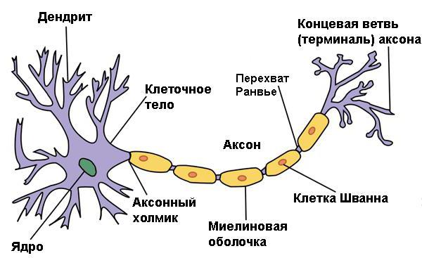
Axons- the main processes of nerve cells, through which information is transmitted along the neural circuit. The more outgoing channels of information transmission a neuron forms, the more branches its axon has.
Dendrites- others On them are located input synapses - specific points where contact with neurons occurs. Therefore, the incoming neural signal is called synoptic transmission.
Classification and properties of nerve cells
Nerve cells, or neurons, are divided into many groups and subgroups, depending on their specialization, functionality, and place in the neural network.
The elements responsible for sensory perception of external stimuli (vision, hearing, tactile sensations, smell, etc.) are called sensory. The neurons that network together to provide motor functions are called motor neurons. Also in the neural network there are mixed neurons that perform universal functions.
Depending on the location of the neuron in relation to the brain and the executive organ, cells can be primary, secondary, etc.
Genetically, neurons are responsible for the synthesis of specific molecules, with the help of which they build synaptic connections with other tissues, but nerve cells do not have the ability to divide.
This is also the basis of the statement, widespread in the literature, that “nerve cells do not recover”. Naturally, neurons unable to divide cannot regenerate. But every second they are able to create many new neural connections to perform complex functions.
Thus, cells are programmed to constantly create more and more new connections. This is how complex communications develop. The creation of new connections in the brain leads to the development of intelligence, thinking. Muscular intelligence also develops in a similar way. The brain is irreversibly improved while learning more and more new motor functions.

The development of emotional intelligence, physical and mental, occurs in the nervous system in a similar way. But if the emphasis is on one thing, other functions develop less rapidly.
Brain
The adult brain weighs approximately 1.3-1.5 kg. Scientists have found that up to 22 years old, its weight gradually increases, and after 75 years it begins to decrease.
There are more than 100 trillion electrical connections in the brain of the average individual, which is several times more than all connections in all electrical devices in the world.
Researchers spend tens of years and tens of millions of dollars on studying and trying to improve brain functions.

Departments of the brain, their functional characteristics
Nevertheless, modern knowledge about the brain can be considered sufficient. Especially considering that the concepts of science about the functions of individual parts of the brain made possible the development of neurology, neurosurgery.
The brain is divided into the following zones:
- Forebrain. The regions of the forebrain are usually ascribed to "higher" mental functions. It includes:
- the frontal lobes, which are responsible for coordinating the functions of other areas;
- those responsible for hearing and speech;
- The parietal lobes regulate movement control and sensory perception.
- the occipital lobes are responsible for visual function.
2. The midbrain includes:
- Thalamus, where almost all information entering the forebrain is processed.
- The hypothalamus controls information from the organs of the central and peripheral nervous system and autonomic NS.
3. The hindbrain includes:

Spinal cord
The average length of the spinal cord in an adult is approximately 44 cm.
It originates from the brain stem and passes through the foramen magnum in the skull. It ends at the level of the second lumbar vertebra. The end of the spinal cord is called the cone of the brain. It ends with an accumulation of lumbar and sacral nerves.
From the spinal cord, 31 pairs of spinal nerves branch out. They help to connect the parts of the nervous system: central and peripheral. Through these processes, parts of the body and internal organs receive signals from the NS.
In the spinal cord, the primary processing of reflex information also takes place, due to which the process of a person's response to stimuli in dangerous situations is accelerated.
CSF, or cerebral fluid, common to the spinal cord and brain, is formed in the vascular nodes of the clefts of the brain from the blood plasma.

Normally, its circulation should be continuous. CSF creates constant internal cranial pressure, performs shock-absorbing and protective functions. Analysis of the composition of the cerebrospinal fluid is one of the simplest ways to diagnose serious NS diseases.
What do lesions of the central nervous system of various origins lead to?
Damage to the nervous system, depending on the period, is divided into:
- Pre-perinatal - brain damage during intrauterine development.
- Perinatal - when the lesion occurs during childbirth and in the first hours after birth.
- Postnatal - when damage to the spinal cord or brain occurs after birth.
Depending on the nature, the lesions of the central nervous system are divided into:
- Traumatic(most obvious). It must be taken into account that the nervous system is of paramount importance for living organisms and from the point of view of evolution, therefore, the spinal cord and brain are reliably protected by a number of membranes, peri-cerebral fluid and bone tissue. However, in some cases, this protection is insufficient. Some injuries cause damage to the central and peripheral nervous systems. Traumatic lesions of the spinal cord more often lead to irreversible consequences. Most often these are paralysis, moreover, degenerative (accompanied by the gradual death of neurons). The higher the damage, the more extensive the paresis (decrease in muscle strength). Open and closed concussions are considered the most common injuries.
- Organic CNS damage often occurs during childbirth and leads to infantile cerebral palsy. They arise due to oxygen starvation (hypoxia). It is the result of prolonged labor or entanglement with the umbilical cord. Depending on the period of hypoxia, cerebral palsy can be of different degrees of severity: from mild to severe, which is accompanied by complex atrophy of the functions of the central and peripheral nervous system. Damage to the central nervous system after stroke is also defined as organic.
- Genetically determined lesions of the central nervous system occur due to mutations in the gene chain. They are considered hereditary. The most common are Down syndrome, Tourette's syndrome, autism (genetic metabolic disorder), which appear immediately after birth or in the first year of life. Diseases of Kensington, Parkinson, Alzheimer are considered degenerative and appear in middle or old age.
- Encephalopathy- most often occur as a result of damage to brain tissue by pathogens (herpetic encephalopathy, meningococcal, cytomegalovirus).

The structure of the peripheral nervous system
The PNS is formed by nerve cells located outside the brain and spinal canal. It consists of (cranial, spinal and vegetative). There are also 31 pairs of nerves and nerve endings in the PNS.
In a functional sense, PNS consists of somatic neurons, which transmit motor impulses and contact with receptors of the sense organs, and vegetative, which are responsible for the activity of internal organs. Peripheral neural structures contain motor, sensory and vegetative fibers.
Inflammatory processes
Diseases of the central and peripheral nervous systems are completely different in nature. While CNS damage most often has complex, global consequences, PNS diseases often manifest themselves in the form of inflammatory processes in the zones of nerve nodes. In medical practice, such inflammation is called neuralgia.
Neuralgia - These are painful inflammations in the area of accumulation of nerve nodes, the irritation of which causes an acute reflexive attack of pain. Neuralgias include polyneuritis, radiculitis, inflammation of the trigeminal or lumbar nerve, plexitis, etc.

The role of the central and peripheral nervous system in the evolution of the human body
The nervous system is the only system of the human body that can be improved. The complex structure of the central and peripheral nervous system of a person is determined genetically and evolutionarily. The brain has a unique property - neuroplasticity. This is the ability of CNS cells to take on the functions of neighboring dead cells, building new neural connections. This explains the medical phenomena when children with organic brain damage develop, learn to walk, speak, etc., and people after a stroke eventually recover the ability to move normally. All this is preceded by the construction of millions of new connections between the central and peripheral parts of the nervous system.
With the progress of various techniques for the recovery of patients after brain injury, techniques for the development of human potential are also being born. They are based on the logical assumption that if both the central and peripheral nervous systems can recover from injury, then healthy nerve cells are also able to develop their potential almost indefinitely.
1. What is related to the peripheral nervous system? How and where are spinal nerves formed and into which branches do they divide?
The peripheral nervous system is that part of the NS that connects GM and SM with sensitive devices - affectors, as well as with those organs and devices that respond to external and internal stimulation with adaptive reactions (movement, secretion of glands) - effectors.
PNS consists of:
Nerves (trunks, plexuses, roots)
Nerve nodes
Peripheral endings
The spinal nerves are formed by the fusion of the posterior and anterior branches, which are anatomically and functionally connected to their segments of the spinal cord through these branches. Therefore, there are 31 pairs of s / m nerves.
The trunk of the s / m nerve is divided into branches:
Anterior branch
Posterior branch
Meningeal branch
White connective vevt
2. Posterior branches of the s / m nerves: their zone of innervation and distribution peculiarity?
The posterior branch has a segmental structure. Therefore, it innervates parts of the body that have retained segmentation: deep muscles of the back, neck, skin over these areas.
The posterior branches are mixed, divided into lateral and medial branches, their diameter is less than the anterior branches. The exceptions are: 1). posterior branch of the 1st cervical s / m nerve (suboccipital nerve) - motor; 2). The posterior branch of the II cervical s / m nerve is sensitive, more than the anterior one.
3. Anterior branches of the s / m nerves: their zone of innervation and difference from the posterior ones?
The anterior branches are not segmented, innervate the parts of the body that have lost their segmentation, form plexuses, the branch is mixed.
4. Why do the anterior branches of the s / m nerves form plexuses? The anterior branches of which nerves do not form them? Why?
ANSWER: plexuses are formed because the anterior branches of the s / m nerves innervate the unsegmented areas. Metamerism is preserved only by the anterior branches of the s / m nerves of Th2 - Th11 segments, they have a segmental structure, they are called intercostal nerves.
5. What plexuses do you know? Their zone of innervation?
Plexus:
· Cervical. From the anterior branches of the 4 upper cervical s / m nerves. Innervates the skin in the neck, diaphragm, neck muscles.
· Shoulder. The anterior branches of the 4 lower cervical s / m nerves. Innervates muscles, skin of the upper extremities, superficial muscles of the chest and back.
· Lumbar plexus. Anterior branches of the lumbar nerves. Innervates the skin, muscles of the lower abdomen, thighs.
· Sacral plexus. Formed by sacral nerves
6. Cranial nerves: how do they differ from spinal nerves and what groups are they divided into according to the fiber composition?
CN - nerves extending from the brain. Differences from s / m nerves:
· They do not have a segmental structure, they are different in function, form, and places of exit.
· Different in composition of fibers.
According to the composition of the fibers, 4 groups are distinguished:
ü Sensitive (1,2,8 pairs of CHN)
ü Motor (3,4,6,11,12 pairs of CHN)
ü Mixed (5,7,9,10 pairs of CHN)
ü Having plus vegetative fibers (3,7,9,10 pairs of CN)
7. What are the peripheral nerves made of? What connective tissue membranes do they have? What is the perineural space, its meaning?
A nerve is a part of the nervous system, which is an elongated cord formed by bundles of nerve fibers and connective tissue sheaths.
They have connective tissue shells of three types:
· Endoneural - m / u with separate nerve fibers, forms separate bundles of nerve fibers;
Perineurium - surrounds several bundles of nerve fibers, formed by two plates:
ü Visceral
ü Parietal
Epineurium - found in the largest nerves, rich in blood vessels - nourishes the nerve, provides collateral circulation.
There is a perineural space between the plates, all CNs have, SMN is controversial, it communicates with the subarachnoid space, contains cerebrospinal fluid. The clinical significance is the advancement of the rabies pathogen through this space to the GM and SM.
8. What is nerve fiber? They are classified according to the caliber and speed of the impulses.
Nerve fiber is a process of a nerve cell surrounded by a sheath of lemmocytes.
By caliber and speed, they are divided into:
· Gr. A: thick myelin fibers up to 100 microns, v = 10-120 m / s, form somatic nerves.
· Gr. B: thin myelin fibers 1-3 microns, v = 3-14 m / s, form pregangliolar autonomic nerves.
· Gr. C: myelin-free fibers 0.4-1.2 microns, v = 0.6-2.4 m / s, form postgangliolar autonomic nerves (to the organs).
9. Intrabarrel structure of nerves.
In addition to the fact that the nerve may include nerve fibers of different f-ii, surrounded by connective tissue sheaths, and having a perineural space, bundles of nerve fibers can be located in different ways. According to Sinelnikov, there are:
· Cable type (vegetative) - all nerve fibers run in parallel;
· Network type (somatic) - adaptive function, a special form of connections with m / u bundles of nerve fibers.
10. Regularities of the location of extraorganic nerves.
· Nerves are paired and diverge symmetrically relative to the central nervous system;
· Nerves reach the organs along the shortest path, with the exception of the nerves of those organs that move in the process of their development, while the nerves lengthen and change their path;
· Nerves innervate the muscles from those segments that correspond to the myotomes of the muscle anlage, if the muscles move, the nerves lengthen.
· Nerves accompany large arteries, veins, forming neurovascular bundles, they are located in protected places.
11. What types of branching of intraorganic nerves depend on? What types of them do you know in muscles with different structure and function?
Muscle innervation options:
· Trunk type - small branches from one large nerve;
Any nerve consists of nerve fibers - a conductive apparatus and sheaths - a supporting connective tissue frame.
Shells
Adventitiy. Adventitium is the densest, most fibrous outer membrane.
Epinsvry. Epineurium is an elastic, elastic connective tissue membrane located under the adventitia.
Perineurium. Perineurium is a covering, consisting of 3-10 layers of epithelioid-type cells, very resistant to stretching, but easily tearing when sutured. The perineurium divides the nerve into bundles containing up to 5000-10000 fibers.
Endoneurium. It is a delicate shell that separates single fibers and small bundles. In this case, it is like a hematoneural barrier.
Peripheral nerves can be considered as a kind of axonal cables, delimited by more or less complex sheaths. These cables are the offshoots of living cells, and the axons themselves are continuously renewed by the flow of molecules. The nerve fibers that make up the nerve are the processes of various neurons. The motor fibers are the processes of the motor neurons of the anterior horns of the spinal cord and the nuclei of the brainstem, the sensitive ones are the dendrites of the pseudo-neurons of the spinal ganglia, the vegetative ones are the axons of the neurons of the border sympathetic trunk.
A separate nerve fiber consists of the actual process of the neuron - the d axial cylinder and the myelin sheath. The myelin sheath is formed by outgrowths of the membrane of Schwann cells and has a phospholipid composition.In this, the peripheral nerve fibers differ from the fibers of the central nervous system. where the myelin sheath is formed by outgrowths of oligodendrocytes.
The blood supply to the nerve is carried out stepwise from adjacent tissues or vessels. A longitudinal vascular network is formed on the surface of the nerve, from which many perforating branches extend to the internal structures of the nerve. With the blood, glucose, oxygen, low-molecular energy substrates enter the nerve fibers, and decay products are removed.
To perform the function of the nerve conduction) "fiber must constantly maintain its structure. However, its own structures performing biosynthesis are not enough to meet the plastic needs in the processes of the neuron. Therefore, the main synthesis occurs in the body of the neuron, followed by the transport of the formed substances along the axon. To a much lesser extent, this the process is carried out by Schwann cells with further transition of metabolites into the axial cylinder of the nerve fiber.
Axonal transport.
The fast and slow modes of movement of substances along the fiber are distinguished.
Fast orthograde axonal transport occurs at a speed of 200-400 mm per day and is mainly responsible for the transfer of membrane constituents: phospholigases, lipoproteins, and membrane enzymes. Retrograde axonal transport ensures the movement of membrane parts in the opposite direction at a speed of up to 150-300 mm per day and their accumulation around the nucleus in close connection with lysosomes. Slow orthograde axonal transport occurs at a rate of 1-4 mm per day and transports soluble proteins and elements of the inner cell scaffold. The volume of substances carried by slow transport is much greater than by fast transport.
Any type of axonal transport is an energy-dependent process performed by contractile proteins analogs of actin and myelin in the presence of macroergs and calcium ions. Energy substrates and ions enter the nerve fiber along with the local blood flow.
Local blood supply to the nerve is an absolute prerequisite for axonal transport.
Neurophysiology of impulse transmission:
Conduction of a nerve impulse along the fiber occurs due to the propagation of a depolarization wave along the sheath of the appendix. Most of the peripheral nerves, along their motor and sensory fibers, provide impulse conduction at a speed of up to 50-60 m / s. The actual depolarization is a rather passive process, while the restoration of the resting membrane potential and the ability to conduct is carried out through the functioning of the NA / K and Ca pumps. For their work, ATP is required, a prerequisite for the formation of which is the presence of segmental blood flow. Cutting off the blood supply to the nerve immediately blocks the conduction of the nerve impulse.
Semiotics of neuropathies
Clinical symptoms that develop with damage to peripheral nerves are determined by the functions of the nerve fibers that form the nerve. According to the three groups of fibers, there are also three groups of symptoms of suffering: motor, sensory and vegetative.
The clinical manifestations of these disorders can be manifested by symptoms of loss of function, which is more common and symptoms of irritation, the latter being a rarer option.
Loss-type motor disorders are manifested by plegias and paresis of a peripheral nature with low tone, low reflexes and hypotrophy. Symptoms of irritation include convulsive contraction of the muscles - krumpy. These are paroxysmal, painful contractions of one or more muscles (what we used to call a cramp). Most often, cramps are localized in the maxillary-hyoid muscle, under the occipital muscle, thigh adductors, quadriceps femoris muscle, triceps calf muscle. The mechanism of the onset of cramps is not clear enough; partial morphological or functional denervation in combination with autonomic irritation is assumed. In this case, the autonomic fibers take on some of the somatic functions and then the striated muscle begins to respond to acetylcholine similarly to smooth muscles.
Sensitive disorders of the type of prolapse are manifested by hypesthesia, anesthesia. The symptoms of irritation are more varied: hyperesthesia, hyperpathy (qualitative perversion of sensation with the acquisition of an unpleasant shade), paresthesia ("chills", burning sensation in the innervation zone), pain along the nerves and roots.
Vegetative disorders are manifested by impaired sweating, suffering from the motor function of hollow internal organs, orthostatic hypotension, trophic changes in the skin and nails. The irritative variant is accompanied by pain with an extremely unpleasant cutting, twisting component, which occurs mainly with damage to the median and tibial nerves, as the richest in vegetative fibers.
It is necessary to pay attention to the variability of the manifestations of neuropathy. Slow changes in the clinical picture occurring over weeks, months really reflect the dynamics of neuropathy, while changes within hours or one or two days are more often associated with changes in blood flow, temperature, electrolyte balance.
Pathophysiology of neuropathy
What happens to nerve fibers in nerve diseases?
There are four main options for changes.
1.Waller's degeneration.
2. Atrophy and degeneration of the axon (axonopathy).
3. Segmental demyelination (myelinopathy).
4. Primary damage to the bodies of nerve cells (neuronopathy).
Wallerian degeneration occurs as a result of gross local damage to the nerve fiber, more often due to mechanical and ischemic factors. The function of conduction along this part of the fiber is completely and immediately disrupted. After 12-24 hours, the structure of the axoplasm changes in the distal part of the fiber, but the conduction of the impulse persists for another 5-6 days. On the 3-5th day, the destruction of the endings of the nerve occurs, and by the 9th day, their disappearance. From 3 to 8 days, the mislinovy membranes are progressively destroyed. In the second week, the division of Schwann cells begins, and by 10-12 days they form longitudinally oriented nerve processes. From 4 to 14 days, multiple growth bulbs appear on the proximal portions of the fibers. The rate of fiber sprouting through the s / t at the site of injury can be extremely low, but distal to the intact parts of the nerve, the rate of regeneration can reach 3-4 mm per day. With this type of lesion, good recovery is possible.
Axonal degeneration occurs as a result of metabolic disturbances in the bodies of neurons, which then causes disease of the processes. The cause of this condition is systemic metabolic diseases and the action of exogenous toxins. Axonal necrosis is accompanied by uptake of myelin and the remains of the axial cylinder by Schwann cells and macrophages. The possibility of restoring nerve function with this suffering is extremely low.
Segmental demyelination is manifested by a primary lesion of the myelin sheaths while the axial cylinder of the fiber is preserved. The severity of the development of impairment may resemble that of mechanical nerve injury, but the impairment of function is easily reversible, sometimes within a few weeks. Pathomorphologically, disproportionately thin myelin sheaths, accumulation of mononuclear phagocytes in the endoneural space, proliferation of Schwann cell processes around the processes of neurons are determined. The restoration of function occurs quickly and in full with the termination of the action of the damaging factor.
Possible karyotypes of Down syndrome
Mitochondrial diseases
The main character traits of a Leo woman
Can i go to the solarium?
How to squeeze out a pimple at home and should you do it?