There is nothing surprising in the fact that the human hearing aid is considered to be the most perfect sensory organ. It contains the highest concentration of nerve cells (over 30,000 sensors).
Human hearing aid
The structure of this apparatus is very complex. People understand the mechanism by which the perception of sounds is carried out, but scientists are not yet fully aware of the sensation of hearing, the essence of signal conversion.
The following main parts are distinguished in the structure of the ear:
- outdoor;
- medium;
- internal.
Each of the above areas is responsible for performing a specific job. The outer part is considered a receiver that receives sounds from the external environment, the middle part is an amplifier, and the inner part is a transmitter.
Human ear structure
The main components of this part:
- ear canal;
- auricle.
The auricle consists of cartilage (it is characterized by firmness, elasticity). From above it is covered with skin. The lobe is located at the bottom. This area has no cartilage. It includes adipose tissue, skin. The auricle is considered a rather sensitive organ.
Anatomy
Smaller elements of the auricle are:
- curl;
- tragus;
- antihelix;
- curl legs;
- antigus.
Koscha is a specific covering lining the ear canal. Inside it contains glands that are considered to be vital. They secrete a secret that protects against many agents (mechanical, thermal, infectious).
The end of the passage is represented by a kind of dead end. This specific barrier (eardrum) is required to separate the outer, middle ear. He begins to vibrate when sound waves hit him. After the sound wave hits the wall, the signal is transmitted further, towards the middle part of the ear.
Blood goes to this site through two branches of the arteries. The outflow of blood is performed through the veins (v. Auricularis posterior, v. Retromandibularis). localized in front, behind the auricle. They also carry out the removal of lymph.
In the photo, the structure of the outer ear
Functions
Let us indicate the significant functions that are assigned to the outer part of the ear. She is capable of:
- take sounds;
- transmit sounds to the middle of the ear;
- direct a wave of sound to the inside of the ear.
Possible pathologies of the disease, injury
Let's note the most common diseases:
The average
The middle ear plays a huge role in signal amplification. Amplification is possible thanks to the auditory ossicles.
Structure
Let's indicate the main components of the middle ear:
- tympanic cavity;
- auditory (eustachian) tube.
The first component (the tympanic membrane) contains a chain inside which small bones are included. The smallest bones play an important role in the transmission of sound vibrations. The eardrum consists of 6 walls. Its cavity contains 3 auditory ossicles:
- hammer. Such a bone is endowed with a rounded head. This is how it connects to the handle;
- anvil. It includes a body, processes (2 pcs.) Of different lengths. Its connection with the stirrup is made by means of a slight oval thickening, which is located at the end of a long process;
- stirrup. In its structure, a small head is distinguished, bearing an articular surface, an anvil, and legs (2 pcs.).
Arteries go to the tympanic cavity from a. carotis externa, being its branches. The lymphatic vessels are directed to the nodes located on the lateral wall of the pharynx, as well as to those nodes that are localized behind the concha.
Middle ear structure

Functions
Chain bones are needed for:
- Conducting sound.
- Vibration transmission.
Muscles located in the middle ear are specialized in various functions:
- protective. Muscle fibers protect the inner ear from sound irritations;
- tonic. Muscle fibers are necessary to maintain the ossicular chain, the tone of the tympanic membrane;
- accommodative. The sound-conducting apparatus adapts to sounds endowed with different characteristics (strength, height).
Pathologies and diseases, trauma
Among the popular diseases of the middle ear, we note:
- (perforated, non-perforated,);
- catarrh of the middle ear.
Acute inflammation can occur with injuries:
- otitis media, mastoiditis;
- otitis media, mastoiditis;
- , mastoiditis, manifested with injuries to the temporal bone.
It can be complicated, uncomplicated. Among the specific inflammations, we point out:
- syphilis;
- tuberculosis;
- exotic diseases.
Anatomy of the outer, middle, inner ear in our video:
Let's point out the significant importance of the vestibular analyzer. It is necessary for the regulation of the position of the body in space, as well as for the regulation of our movements.
Anatomy
The periphery of the vestibular analyzer is considered to be the area of the inner ear. In its composition, we highlight:
- semicircular canals (these parts are located in 3 planes);
- statocyst organs (they are represented by sacs: oval, round).
The planes are called: horizontal, frontal, sagittal. The two pouches represent the vestibule. A round pouch is found near the curl. The oval sac is placed closer to the semicircular canals.
Functions
Initially, the analyzer is excited. Then, thanks to the vestibulo-spinal nerve connections, somatic reactions occur. Such reactions are needed to redistribute muscle tone, maintain body balance in space.
The connection between the vestibular nuclei, the cerebellum determines the mobile reactions, as well as all the reactions for the coordination of movements that appear when performing sports, labor exercises. To maintain balance, vision, muscular-articular innervation are very important.

The ear is the organ of hearing and balance. The ear is located in the temporal bone and is conditionally divided into three sections: external, middle and internal.
Outer ear formed by the auricle and the external auditory canal. The border between the outer and middle ear is eardrum.
The auricle is formed by three tissues:
a thin plate of hyaline cartilage, covered on both sides by the perichondrium, having a complex convex-concave shape, which determines the relief of the auricle;
skin very thin, tight to the perichondrium and almost no fatty tissue;
subcutaneous fatty tissue located in significant numbers in the lower part of the auricle.
The following elements of the auricle are usually distinguished:
curl- free upper-outer edge of the shell;
antihelix- an elevation running parallel to the curl;
tragus- a protruding section of cartilage located in front of the external auditory canal and being part of it;
antigus- a ledge located posterior to the tragus and the notch separating them;
lobe, or a lobule, of the ear, devoid of cartilage and consisting of fatty tissue covered with skin. The auricle is attached to the temporal bone by rudimentary muscles. The anatomical structure of the auricle determines the features of the pathological processes that develop during trauma, with the formation of an otogematoma and perichondritis.
Sometimes there is a congenital underdevelopment of the auricle - microtia or its complete absence of anotia.
External auditory canal is a canal that begins with a funnel-shaped depression on the surface of the auricle and is directed in an adult horizontally from front to back and from bottom to top to the border with the middle ear.
There are the following sections of the external auditory canal: external membranous cartilaginous and internal - bone.
External membranous cartilage takes 2/3 of the length. In this section, the front and lower walls are formed by the cartilaginous tissue, and the back and upper walls have fibro-connective tissue.
Anterior wall of the external auditory canal borders on the joint of the lower jaw, and therefore the inflammatory process in this area is accompanied by sharp pain when chewing.
Top wall separates the outer ear from the middle cranial fossa, therefore, with fractures of the base of the skull, cerebrospinal fluid with an admixture of blood flows out of the ear. The cartilaginous plate of the external auditory canal is interrupted by two transverse slits, which are closed by fibrous tissue. Placing them next to the salivary gland can help spread the infection from the outer ear to the salivary gland and the mandibular joint.
The skin of the cartilaginous section contains a large number of hair follicles, sebaceous and sulfur glands. The latter are modified sebaceous glands that secrete a special secret, which, together with the secreted sebaceous glands and the detached skin epithelium, forms earwax. The removal of dried sulfur plates is facilitated by vibrations of the membranous-cartilaginous part of the external auditory canal during chewing. The presence of abundant grease in the outer part of the ear canal prevents water from entering it. There is a tendency for the ear canal to narrow from the entrance to the end of the cartilaginous portion. Attempts to remove sulfur using foreign objects can lead to pushing pieces of sulfur into the bone section, from where it cannot be evacuated on its own. Conditions are created for the formation of a sulfur plug and the development of inflammatory processes in the outer ear.
Internal bony part of the ear canal has in its middle the narrowest place - an isthmus, behind which a wider section is located. Unskillful attempts to remove a foreign body from the ear canal can lead to pushing it behind the isthmus, which greatly complicates further removal. The skin of the bone section is thin, does not contain hair follicles and glands, and passes to the tympanic membrane, forming its outer layer.
The middle ear consists of the following elements: the tympanic membrane, the tympanic cavity, the ossicles, the auditory tube and the air cells of the mastoid process.
Eardrum is the boundary between the outer and middle ear and is a thin, pearlescent gray membrane impermeable to air and liquid. Most of the tympanic membrane is in a taut state due to fixation in the circular groove of the fibrocartilaginous ring. In the upper anterior section, the tympanic membrane is not stretched due to the absence of the groove and the middle fibrous layer.
The tympanic membrane consists of three layers:
1 - external - cutaneous is a continuation of the skin of the external auditory canal, is thinned and does not contain glands and hair follicles;
2 - internal - slimy- is a continuation of the mucous membrane of the tympanic cavity;
3 - medium - connective tissue- represented by two layers of fibers (radial and circular), providing a taut position of the tympanic membrane. When it is damaged, a scar is usually formed due to the regeneration of the skin and mucous layer.
Otoscopy - examination of the tympanic membrane is of great importance in the diagnosis of ear diseases, as it gives an idea of the processes taking place in the tympanic cavity. Tympanic cavity is a cube of irregular shape with a volume of about 1 cm3, located in the petrous part of the temporal bone. The tympanic cavity is divided into 3 sections:
1 - upper - attic, or the eardrum space (epitympanum), located above the level of the tympanic membrane;
2 - medium - (mesotympanum) located at the level of the stretched part of the tympanic membrane;
3 - lower - (hypotympanum) located below the level of the tympanic membrane and passing into the auditory tube.
The tympanic cavity has six walls, which are lined with mucous membrane, supplied with ciliated epithelium.
1 - outer wall represented by the tympanic membrane and bony parts of the external auditory canal;
2 - inner wall is the border of the middle and inner ear and has two openings: the window of the vestibule and the window of the cochlea, closed by the secondary tympanic membrane;
3 - upper wall (roof of the tympanic cavity)- is a thin bone plate that borders the middle cranial fossa and the temporal lobe of the brain;
4 - lower wall (bottom of the tympanic cavity)- bordered by the bulb of the jugular vein;
5 - front wall borders on the internal carotid artery and in the lower section has the mouth of the auditory tube;
6 - back wall- separates the tympanic cavity from the air cells of the mastoid process and communicates with them in the upper part through the entrance to the mastoid process cave.
Auditory bones represent a single chain from the tympanic membrane to the oval window of the vestibule. They are suspended in the tympanic space with the help of connective tissue fibers, covered with a mucous membrane and have the following names:
1 - hammer the handle of which is connected to the fibrous layer of the tympanic membrane;
2 - anvil- occupies a middle position and is connected by joints with the rest of the bones;
3 - stirrup, the footplate of which transmits vibrations in the vestibule of the inner ear.
Muscles of the tympanic cavity(stretching the eardrum and stapes) keep the ossicles in a state of tension and protect the inner ear from excessive sound stimuli.
Auditory tube- education 3.5 cm long, through which the tympanic cavity communicates with the nasopharynx. The auditory tube consists of a short bony section, which occupies 1/3 of the length, and a long membranous-cartilaginous section, which is a closed muscular tube that opens when swallowing and yawning. The junction of these departments is the narrowest and is called the isthmus.
The mucous membrane lining the auditory tube, is a continuation of the mucous membrane of the nasopharynx, covered with multi-row cylindrical ciliated epithelium with the movement of cilia from the tympanic cavity to the nasopharynx. Thus, the auditory tube performs a protective function, preventing the penetration of the infectious agent, and a drainage function, evacuating the discharge from the tympanic cavity. Another important function of the auditory tube is ventilation, which ensures the passage of air and balances atmospheric pressure with the pressure in the tympanic cavity. If the patency of the auditory tube is impaired, air is discharged in the middle ear, the tympanic membrane is drawn in, and persistent hearing loss may develop.
Mastoid cells are air cavities associated with the tympanic cavity in the attic through the entrance to the cave. The mucous membrane lining the cells is a continuation of the mucous membrane of the tympanic cavity.
Internal structure of the mastoid process depends on the formation of air cavities and is of three types:
pneumatic- (most often) - with a large number of air cells;
diploetic- (spongy) - has a few small cells;
sclerotic- (compact) - the mastoid process is formed by dense tissue.
The process of pneumatization of the mastoid process is influenced by past diseases, metabolic disorders. Chronic inflammation of the middle ear can contribute to the development of the sclerotic mastoid process.
All air cavities, regardless of their structure, communicate with each other and the cave - a permanently existing cell. It is usually located at a depth of about 2 cm from the surface of the mastoid process and borders on the dura mater, the sigmoid sinus, and the bony canal in which the facial nerve passes. Therefore, acute and chronic inflammation of the middle ear can lead to the penetration of infection into the cranial cavity, the development of facial nerve paralysis.
Features of the structure of the ear in young children
Anatomical, physiological and immunobiological characteristics of the child's body determine the characteristics of the clinical course of ear diseases in young children. This is reflected in the frequency of inflammatory diseases of the middle ear, the severity of the course, more frequent complications, the transition of the process to a chronic one. Ear diseases in early childhood contribute to the development of complications in older children and in adulthood. Anatomical and physiological features of the ear in young children occur in all departments.
Auricle in a nursing child, it is soft, low elastic. The curl and lobe are not distinctly expressed. The auricle is formed by the age of four.
External auditory canal in a newborn child, it is short, it is a narrow gap filled with original lubricant. The bony part of the wall is not yet developed and the upper wall is adjacent to the lower one. The ear canal is directed forward and downward, therefore, in order to inspect the ear canal, the auricle must be pulled back and downward.
Eardrum denser than in adults due to the outer skin layer, which has not yet formed. In this regard, in acute otitis media, perforation of the tympanic membrane occurs less frequently, which contributes to the development of complications.
Tympanic cavity in newborns, it is filled with myxoid tissue, which is a good breeding ground for microorganisms, and therefore the risk of developing otitis media at this age increases. Resorption of myxoid tissue begins from 2-3 weeks of age, however, it can be in the tympanic cavity during the first year of life.
Auditory tube at an early age, it is short, wide and horizontally located, which facilitates easy penetration of infection from the nasopharynx into the middle ear.
Mastoid does not have formed air cells, except for the cave (antrum), which is located directly under the outer surface of the mastoid process in the area of the Shipo triangle. Therefore, in the inflammatory process (antritis), a painful infiltrate with protrusion of the auricle often develops in the behind-the-ear region. In the absence of the necessary treatment, intracranial complications are possible. Pneumatization of the mastoid process occurs as the child grows and ends at the age of 25-30.
Temporal bone in a newborn child, it consists of three independent elements: scales, mastoid process and pyramid due to the fact that they are separated by cartilaginous growth zones. In addition, birth defects are often found in the temporal bone, which contribute to the more frequent development of intracranial complications.
The inner ear is represented by a bony labyrinth located in the pyramid of the temporal bone, and a membranous labyrinth located in it.
The bony labyrinth consists of three sections: vestibule, cochlea, and three semicircular canals.
The vestibule is the middle part of the maze, on the outer wall of which there are two windows leading to the tympanic cavity. Oval window the vestibule is closed with a stirrup plate. Round window closed by a secondary tympanic membrane. The front part of the vestibule communicates with the snail through the vestibule stairs. The back part contains two indentations for the sacs of the vestibular apparatus.
Snail- a bony spiral canal in two and a half turns, which is divided by a bony spiral plate into the staircase of the vestibule and the tympanic staircase. They communicate with each other through a hole located at the top of the snail.
Semicircular canals- bone formations located in three mutually perpendicular planes: horizontal, frontal and sagittal. Each canal has two knees - an extended leg (ampulla) and a simple one. The simple legs of the anterior and posterior semicircular canals merge into one, so the three canals have five openings.
Webbed labyrinth consists of a membranous cochlea, three semicircular canals and two sacs (spherical and elliptical) located on the eve of the bone labyrinth. Between the bone and membranous labyrinth there is perilymph, which is a modified cerebrospinal fluid. The membranous labyrinth is filled endolymph.
In the inner ear there are two analyzers connected anatomically and functionally - auditory and vestibular. Auditory analyzer located in the cochlear duct. A vestibular- in three semicircular canals and two sacs of the vestibule.
Auditory peripheral analyzer. In the upper corridor of the snail is located coiled (Corti's) organ, which is the peripheral part of the auditory analyzer. In section, it has a triangular shape. Its lower wall is the main membrane. Above is the vestibule (Reissner) membrane. The outer wall is formed by a spiral ligament and cells of the vascular stria located on it.
The main membrane consists of elastic elastic transverse fibers stretched in the form of strings. Their length increases from the base of the cochlea to the apex. The spiral (Corti) organ has a very complex structure and consists of inner and outer rows of sensitive hairy bipolar cells and supporting (supporting) cells. The processes of the hair cells of the spiral organ (auditory hairs) come into contact with the integumentary membrane and, when the main plate vibrates, they are irritated, as a result of which mechanical energy is transformed into a nerve impulse that spreads to the spiral ganglion, then along the VIII pair of cranial nerves to the medulla oblongata. Subsequently, most of the fibers pass to the opposite side and along the conducting paths the impulse is transmitted to the cortical part of the auditory analyzer - the temporal lobe of the hemisphere.
Vestibular peripheral analyzer. On the eve of the labyrinth, there are two membranous sacs with an otolith apparatus located in them. On the inner surface of the sacs there are elevations (spots) lined with neuroepithelium, consisting of supporting and hair cells. The hairs of sensitive cells form a network that is covered with a jelly-like substance containing microscopic crystals - otoliths. With rectilinear body movements, the otoliths are displaced and mechanical pressure, which causes irritation of the neuroepithelial cells. The impulse is transmitted to the vestibular node, and then along the vestibular nerve (VIII pair) to the medulla oblongata.
On the inner surface of the ampullae of the membranous ducts there is a protrusion - an ampullar ridge, consisting of sensitive neuroepithelial cells and supporting cells. Sensitive hairs sticking together are presented in the form of a brush (cupula). Irritation of the neuroepithelium occurs as a result of movement of the endolymph when the body is displaced at an angle (angular acceleration). The impulse is transmitted by the fibers of the vestibular branch of the vestibular cochlear nerve, which ends in the nuclei of the medulla oblongata. This vestibular area is associated with the cerebellum, spinal cord, nuclei of the oculomotor centers, and the cerebral cortex.
The outer ear consists of the auricle and the external auditory canal.
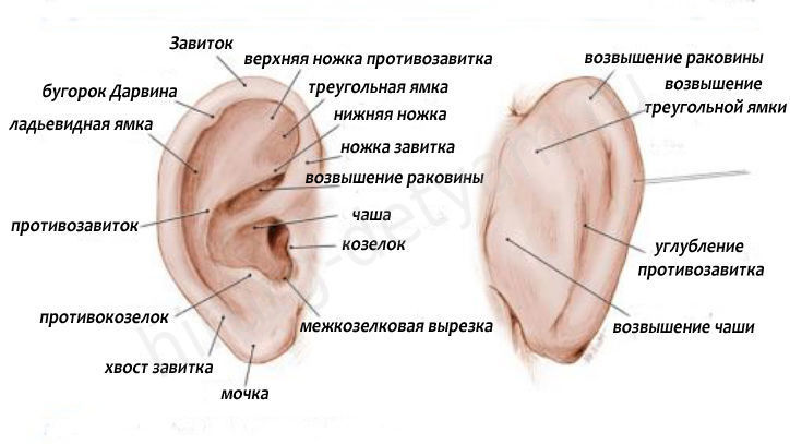
Auricle(auricula) has a complex configuration and is divided into two sections: a lobe, which is a duplication of skin with fatty tissue inside, and a part consisting of cartilage covered with thin skin. If it is possible to fold the skin on the back surface, then on the front surface this cannot be done due to the firm fusion of the skin with the perichondrium. The auricle has a helix, an anthelix, a tragus, an antitragus, and a lobulus. The tragus covers the entrance to the external auditory canal (Fig. 151).
Rice. 151. The structure of the auricle.
1.Triangular fossa; 2. Rook; 3.Legs antihelix; 4.Furl leg; 5. Curl; 6. Sink cavity; 7.Anti-curl; 8. Tragus; 9.Anti-grouse. 10. Lobe.
Pressure on the tragus area can be painful with an inflammatory process in the external auditory canal, and in children and with acute otitis media, since in early childhood the external auditory canal does not have a bony section and therefore it is shorter. Pressure on the tragus in these cases leads, in fact, to pressure on the inflamed tympanic membrane, which is accompanied by increased pain. In addition to these protrusions on the front surface of the auricle, there are depressions - a triangular fossa (fossa triangularis), a boat (scapha). It is necessary to know about these elements of the auricle in order to localize certain processes in the area of the auricle: hematoma in the area of the triangular fossa, abscess of the lobe, etc. It is believed that the height of the auricle normally corresponds to the length of the nasal dorsum. A deviation in one direction or another allows us to speak of microtia or macrotia. The fact that the auricle is distant from the surface of the skull and has features of blood supply (on the anterior surface of the auricle, the vessels are not surrounded by subcutaneous tissue) creates conditions for frostbite, since the vessels are in a state of spasm under the influence of cold. The auricle plays an important role in ototope, i.e. the ability to determine the direction of the sound source, has a protective function. A normal auricle, due to its complex profile, contributes to the retention of dust particles in the outermost part of the ear canal. With a deformed shell, or completely lost, dust reaches the tympanic membrane and, deposited on it, can contribute to the development of inflammation. The auricle to a certain extent affects the acuity of hearing, therefore, to perceive a weak sound, a person puts his hand to the auricle, as if increasing its area.
The auricle, narrowing in a funnel shape, passes into external auditory canal, consisting of two departments: external membranous cartilaginous and internal bone(152) . The diameter of the external auditory canal varies, but this does not affect hearing acuity. In children of the first year of life, the bony part of the external auditory canal is absent, and only the cartilaginous one exists. The length of the external auditory canal in children is 0.5-0.7 cm, in an adult it is about 3 cm.
Fig. 152. External auditory canal.
The cartilaginous part of the auditory canal, consisting partly of cartilaginous tissue, is bordered from below by the capsule of the parotid salivary gland. The lower wall has several transverse cracks in the cartilage tissue. Through them, the inflammatory process can spread to the parotid gland. In the cartilaginous section, there are many glands that produce earwax, as well as hair with hair follicles, which can become inflamed when pathogenic flora penetrates, resulting in a furuncle of the external auditory canal.
The cartilaginous section of the external auditory canal is represented by a cartilage groove. This groove is open in the region of the posterior - upper wall and therefore the incisions of the ear canal during surgical interventions on the ear, in order to avoid the occurrence of perichondritis, should be carried out precisely along the posterior - upper wall.
The anterior wall of the external auditory canal is closely adjacent to the temporomandibular joint, and with each chewing movement, this wall moves. In cases where a boil develops on this wall, each chewing movement increases the pain. The close contact of the external auditory canal with the temporomandibular joint causes a fracture of the anterior wall of the auditory canal upon impact in the chin area with rupture of the skin and possible cicatricial obliteration of the lumen of the auditory canal. In addition, the close anatomical relationship of these formations explains the occurrence of some syndromes related to otorhinolaryngology and dentistry. The bony section of the external auditory canal is lined with thin skin, there is a narrowing on the border with the cartilaginous section. Pushing foreign bodies behind this narrowing makes it much more difficult to remove them by one method or another.
The upper wall of the bony section is bordered by the middle cranial fossa, the posterior wall - with the air cells of the mastoid process and, in particular, with the cave. This circumstance explains the occurrence of one of the pathognomonic symptoms of an acute inflammatory process in the mastoid process (mastoiditis) - a symptom of overhanging the posterior - upper wall in the bony part of the ear canal, which leads to a narrowing of its lumen due to developing periostitis.
The skin of the external auditory canal in the cartilaginous part is supplied with hair, sebaceous and sulfur glands. The latter secrete sulfur and are modified sebaceous glands. In the bony part of the external auditory canal, the skin is thin and devoid of hair and glands.
Blood supply the outer ear is provided by the branches of the external carotid artery. The auricle is supplied with blood from posterior auricular and superficial temporal arteries(a. auricularis posterior et a.temporalis superficialis). The same vessels, as well as the deep ear artery ( a.auricularis profunda) provide blood to the deeper sections and the tympanic membrane, form a plexus around the external auditory canal.
Venous outflow is carried out anteriorly in posterosmandibular vein(v. retromandibularis) and posterior to the posterior ear vein (v. auricularis posterior).
Innervation of the outer ear(the skin of the external auditory canal, auricle) is carried out from the third branch of the trigeminal, vagus and glossopharyngeal nerves. This causes the occurrence of "reflected" pain, for example, with inflammation of the periodontal tissues of the eighth lower tooth, a sensation of severe pain in the ear on the corresponding side is possible.
Ear - paired ( right and left), a symmetrical, complex organ of balance and hearing.
Anatomically, the ear is divided into three parts.
#1. Outer ear represented by the external auditory canal, the length of which is 30 mm, as well as the auricle, the basis of which is elastic cartilage 1 mm thick. From above, the cartilage is covered with perichondrium and skin. The lower part of the shell is a lobe. It is devoid of cartilage and is formed by fatty tissue, which is also covered by the skin. Almost every little girl is punctured by parents ( in other words - piercing) the lobes of each ear and decorate them with earrings. Ears should be pierced in compliance with the rules of asepsis to avoid local and general infection.
The free edge of the ear concha forms a curl. Parallel to the curl is the antihelix, in front of which is the cavity of the ear concha. In the ear, a tragus and an antigus are also distinguished. The auricle is attached to the mastoid and zygomatic processes, as well as the temporal bone with the help of muscles and ligaments. The human ear is inactive due to the fact that the muscles that rotate it are practically atrophied. The entrance to the outer ear is covered with hair and contains sebaceous glands. The shape of the auricles, like fingerprints, is individual for all people.
The ear canal connects the auricle and the eardrum. In adults it is longer and narrower, and in children it is shorter and wider. That is why otitis media is more common in early childhood. The skin of the ear canal contains sulfur and sebaceous glands.
# 2. Middle ear represented by the tympanic cavity, which is located in the temporal bone. It contains the smallest auditory ossicles in the human body: the hammer, stapes and incus. With their help, sound is transmitted to the inner ear. The Eustachian tube connects the middle ear cavity with the nasopharynx;
# 3. Inner ear the most complex in structure of all parts. It communicates with the middle ear through a round and oval window. Another name for the inner ear is the membranous labyrinth. It is immersed inside the bone labyrinth. It includes:
the cochlea is the direct organ of hearing;
vestibule and semicircular tubules - are responsible for acceleration, body position in space and balance.
Basic functions of the ear
Perceives sound vibrations;provides balance and position of the human body in space.
Embryonic ear development
Starting from the 4th week of embryonic development, the rudiments of the inner ear are formed. Initially, it is represented by a limited section of the ectoderm. The entire inner ear is formed by the 9th week of intrauterine life. The middle and outer ear are formed from the branchial slits starting from the 5th week. In a newborn, the tympanic cavity is fully formed, the lumen of which is filled with myxoidal tissue. It is absorbed only by the 6th month of a child's life and is a good breeding ground for bacteria.Ear diseases
Among the common pathologies of the ear are distinguished: trauma ( barotrauma, acoustic trauma, etc.), congenital developmental anomalies, diseases ( otitis media, labyrinthitis, etc.).#1. Barotrauma- Damage to the paranasal sinuses of the ear or the Eustachian tube due to changes in ambient pressure. Reasons: flying in an airplane, diving, etc. At the time of injury, there is severe pain, congestion and a feeling of a strong blow. Hearing loss, ringing and tinnitus are immediately noted. A ruptured eardrum is accompanied by bleeding from the ear canal;
# 2. Congenital anomalies ears occur in the first 4 months of intrauterine development due to genetic defects. Ear abnormalities are often associated with malformations of the face and skull. Frequent pathologies: absence of ears, macrotia - excessively large ears, microtia - very small ears. Pathologies of development of the middle ear include: underdevelopment of the auditory ossicles, infection of the inner ear, etc.;
#3. The most common ear condition between 2 and 8 years of age is otitis media... This is due to the anatomical features of the ear. You can guess that a small child's ear hurts if you press on the tragus. Usually the child starts to worry and cry. Characteristic signs of the disease: shooting pain, which can radiate to the head, and intensify when swallowing, sneezing. The common cold contributes to the disease. As a rule, otitis media is combined with rhinitis and tonsillitis;
#4. Labyrinthitis- internal otitis media. It occurs due to not fully treated otitis media. Sometimes the infection "rises" from the teeth affected by caries by the hematogenous route. Symptoms of the disease: hearing loss, nystagmus ( involuntary movement of the eyeball) on the affected side, nausea, tinnitus, etc.
Diagnostics
The definition of the disease begins with a survey and examination of the patient by a doctor. During examination of the auditory opening in adults, the concha is pulled back and upward, and in children - backward and downward. Pulling straightens the ear canal and makes it possible to view it with the help of the auditory funnel to the bony part. During palpation, the doctor presses on the tragus, the cause of the pain in which indicates an inflammation of the middle ear. In addition, the doctor pays attention to regional lymph nodes, which are not normally detected. The eardrum is examined using an otoscope.Instrumental research methods:
X-ray of the temporal bone is of great importance for the diagnosis of various pathological formations of the middle and inner ear;
MRI allows you to get more detailed information about the pathology of the ear, it is especially often used to diagnose tumor and inflammatory changes.
Treatment
A doctor - an otolaryngologist is involved in the treatment of diseases of the ears, as well as the throat and nose.The most common dosage form used to treat ear conditions is drops. With their help, local diseases of the outer and middle ear are treated. If the pathological process has affected the inner ear, as well as nearby organs ( nose, throat, etc.), then general action drugs are prescribed ( antibiotics, pain relievers, etc.). In some advanced cases, for example, with fistular labyrinthitis, surgery is performed.
How to remove sulfur plug? Sulfur is an important substance secreted by the glands of the outer ear. It performs a protective function, always standing out towards the external auditory canal. As a rule, sulfur plugs occur in persons who too often or, conversely, very rarely clean their ears. The most common sign of cerumen is ear congestion. In addition, some people have itchy ears with sulfur plugs. You can try to remove the sulfur plug at home. To do this, you need to drip a warm solution of hydrogen peroxide into the ear. The sulfuric plug will dissolve and the hearing will be restored. In a polyclinic, the ear is washed with warm water using Janet's syringe.
Ear transplant
A person who has lost an ear, for example, in a car accident, has a chance to regain a new, identical organ. Currently, this is done through the cultivation of auricles. For the first time the ear was grown in the laboratories of America. To grow the new organ, it took a mouse to inject ear cartilage cells into its back. The body has successfully accepted an implant grown in this way. Currently, hundreds of such operations are performed in the United States. A cheaper option to replace the auricle is prosthetics. The artificial ear prosthesis is made of hypoallergenic silicone. Such operations, restoring the normal image of a person's face after emergencies, are performed in all countries of the world. For babies with no ears, Cornell doctors and biomedical scientists create auricles using injection dies and 3-D printing. In case of congenital pathology of the middle ear, in particular in the absence or underdevelopment of the auditory ossicles, implantation of a bone conduction hearing aid is performed.Prevention of ear diseases
To prevent the ingress of water before bathing, you must use special tampons for the ears;while bathing the child, avoid getting wet by keeping the head above the water. After feeding, you should keep the baby upright for 5 - 10 minutes so that the air comes out and food does not get into the nasopharynx;
in order to avoid the formation of sulfur plugs, as well as mechanical injury, it is not recommended to clean your ears often using sharp objects. You should clean the auricle with warm water, soap with your fingers;
you should avoid activities that promote the entry of a foreign body into the ear.
The ear is the organ of hearing and balance. The ear is located in the temporal bone and is conditionally divided into three sections: external, middle and internal.
Outer ear educated auricle and external auditory canal. The boundary between the outer and middle ear is the eardrum.
Auricle formed by three fabrics:
Thin plate hyaline cartilage, covered on both sides perichondrium, having a complex convex-concave shape, which determines the relief of the ear
shells;
- skin very thin, tight to the perichondrium and almost no fatty tissue;
- subcutaneous fatty tissue, located in significant numbers in the lower part of the auricle.
The following elements of the auricle are usually distinguished:
- curl- the free top is the non-outer edge of the shell;
- antihelix- an elevation running parallel to the curl;
- tragus- a protruding section of cartilage located in front of the external auditory canal and being part of it;
- antigus - protrusion posterior to
tragus and cuttings separating them;
- lobe, or lobule, ear, devoid of cartilage and consisting of fatty tissue covered with skin.
The auricle is attached to the temporal bone by rudimentary muscles. The anatomical structure of the auricle determines the features of the pathological processes that develop during trauma, with the formation of an otogematoma and perichondritis.
Occasionally occurs congenital underdevelopment auricle - microtia or its complete absence anotia. In such cases, a cosmetic operation is performed, an auricle plastic is formed from a skin fold using a frame made of preserved cartilage or artificial materials. Congenital pathology of the auricle is often combined with developmental anomalies in other parts of the ear - overgrowth of the external auditory canal, malformations of the middle and inner ear.
External auditory canal is a canal that begins with a funnel-shaped depression on the surface of the auricle and is directed in an adult horizontally from front to back and from bottom to top to the border with the middle ear. Therefore, to align the passage during examination, it is necessary to pull the auricle back and up.
There are the following sections of the external auditory canal: external membranous cartilaginous and internal- bone.
External membranous cartilage takes 2/3 of the length. In this section, the front and lower walls are formed by the cartilaginous tissue, and the back and upper walls have fibro-connective tissue.
The anterior wall of the external auditory canal borders on the joint of the lower jaw, and therefore the inflammatory process in this area is accompanied by sharp pain when chewing.
The upper wall separates the outer ear from the middle cranial fossa, therefore, with fractures of the base of the skull, cerebrospinal fluid with an admixture of blood flows out of the ear. The cartilaginous plate of the external auditory canal is interrupted by two transverse slits, which are closed by fibrous tissue. Placing them next to the salivary gland can help spread the infection from the outer ear to the salivary gland and the mandibular joint.
Leather the cartilaginous section contains a large number of hair follicles, sebaceous and sulfur glands. The latter are modified sebaceous glands that secrete a special secret, which, together with the secreted sebaceous glands and the detached skin epithelium, forms earwax. The removal of dried sulfur plates is facilitated by vibrations of the membranous-cartilaginous part of the external auditory canal during chewing. The presence of abundant grease in the outer part of the ear canal prevents water from entering it. There is a tendency for the ear canal to narrow from Echod to the end of the cartilaginous part. Attempts to remove sulfur with the help of foreign objects can lead to pushing pieces of sulfur into the bone section, from where it cannot be evacuated on its own. Conditions are created for the formation of a sulfur plug and the development of inflammatory processes in the outer ear. Therefore, personal ear hygiene should be limited to rinsing the entrance to the ear canal with warm water and soap.
Internal bone section the ear canal has the narrowest point in its middle - isthmus, behind which is a wider area. Unskillful attempts to remove a foreign body from the ear canal can lead to pushing it behind the isthmus, which greatly complicates further removal. The skin of the bone section is thin, does not contain hair follicles and glands and passes to the eardrum, forming its outer layer.
Blood supply to the outer ear provided by the external carotid artery. Venous outflow is carried out into the posterior facial veins.
Lymphatic drainage occurs in the lymph nodes in front of the tragus and under the lower wall of the external auditory canal, as well as in the deep lymph nodes of the neck.
Innervation of the outer ear carried out by the third branch of the trigeminal nerve, the facial nerve, as well as the branch of the vagus nerve, which explains the cough and discomfort when manipulating the ear canal or moving a foreign body in it.
The middle ear is made up of the following elements: tympanic membrane, tympanic cavity, auditory ossicles, auditory tube and air cells of the mastoid process.
Eardrum is the boundary between the outer and middle ear and is a thin, pearlescent-gray membrane impermeable to air and liquid. Most of the tympanic membrane is in a taut state due to fixation in the circular groove of the fibrocartilaginous ring. In the upper anterior section, the tympanic membrane is not stretched due to the absence of the groove and the middle fibrous layer.
The tympanic recirculation consists of three layers: x X - external - cutaneous is a continuation of the skin of the external auditory canal, is thinned and does not contain glands and hair follicles; and 2 - interior- slimy - is a continuation of the mucous membrane of the tympanic cavity; f s - average- connective tissue - is represented by two layers of fibers (radial and circular), providing a taut position of the tympanic membrane. When it is damaged, a scar is usually formed due to the regeneration of the skin and mucous layer.
Otoscopy- examination of the tympanic membrane is of great importance in the diagnosis of ear diseases, as it gives an idea of the processes occurring in the tympanic cavity. Normally, when examining the tympanic membrane, a pelamutric gray color and pronounced identifying signs:
* 1 - short process of the malleus, situated
on the border of the stretched and relaxed part of the tympanic membrane;
* 2 - hammer handle, going from the short process to the center of the tympanic membrane;
* 3 - light cone - shiny triangle with ver
a splint in the center of the tympanic membrane and the base at the edge of it. It is the result of the reflection of light from the frontal reflector and is noted only when the eardrum is in the correct position.
Tympanic cavity is a cube of irregular shape with a volume of about 1 cm 3, located in the petrous part of the temporal bone. The tympanic cavity is divided into 3 sections:
* 1 - upper - attic, or drum space (epitijpanum), located above the level of the tympanic membrane;
* 2 - average- (mesotympanum) located at the level of the stretched part of the tympanic membrane;
* 3 - lower- (hypotympanum), located below the level of the tympanic membrane and passing into the auditory tube.
The tympanic cavity has six walls, which are lined with mucous membrane supplied with ciliated epithelium.
* 1 - the outer wall is represented by the tympanic membrane and the bony parts of the external auditory canal;
* 2 - the inner wall is the border of the middle and inner ear and has two openings: the window of the vestibule and the window of the cochlea, closed by the secondary tympanic membrane;
* 3 - the upper wall (roof of the tympanic cavity) - is a thin bone plate that borders on the middle cranial fossa and the temporal lobe of the brain;
* 4 - the lower wall (bottom of the tympanic cavity) - borders on the bulb of the jugular vein;
* 5 - the anterior wall borders on the internal carotid artery and has the mouth of the auditory tube in the lower section;
* 6 - back wall - separates the tympanic cavity from the air cells of the mastoid process and communicates with them in the upper part through the entrance to the mastoid process cave.
Auditory bones represent a single chain from the tympanic membrane to the oval window of the vestibule. They are suspended in the tympanic space with the help of connective tissue fibers, covered with a mucous membrane and have the following names:
* 1 - hammer, the handle of which is connected to the fibrous layer of the tympanic membrane;
* 2 - anvil- occupies a middle position and is connected by joints with the rest of the bones;
* 3 - stapes, the footplate of which transmits vibrations to the vestibule of the inner ear. Muscle the tympanic cavity (stretching the eardrum and stapedius) keeps the auditory ossicles in a state of tension and protects the inner ear from excessive sound stimuli.
Auditory pipe- education 3.5 cm long, through which the tympanic cavity communicates with the nasopharynx. The auditory tube consists of a short bone department, occupying 1/3 of the length, and long membranous-cartilaginous department, representing a closed muscle tube that opens up when swallowing and yawning. The junction of these departments is the narrowest and is called isthmus.
The mucous membrane lining the auditory tube is a continuation of the nasopharyngeal mucosa, covered with multi-row cylindrical ciliated epithelium with the movement of cilia from the tympanic cavity into the nasopharynx. Thus, the auditory tube performs protective function, preventing the penetration of the infectious agent, and drainage function, evacuating the discharge from the tympanic cavity. Another important function of the auditory tube is ventilation, which ensures the passage of air and balances atmospheric pressure with the pressure in the tympanic cavity. If the patency of the auditory tube is impaired, air is discharged in the middle ear, the tympanic membrane is drawn in, and persistent hearing loss may develop.
Cells mastoid are air cavities associated with the tympanic cavity in the attic through the entrance to the cave. The mucous membrane lining the cells is a continuation of the mucous membrane of the tympanic cavity.
The internal structure of the mastoid process depends on the formation of air cavities, there are three types:
Pneumatic - (most often) - with a large number of air cells;
Diploetic - (spongy) - has few cells
small size;
Sclerotic - (compact) - the mastoid process is formed by dense tissue.
The process of pneumatization of the mastoid process is influenced by past diseases, metabolic disorders. Chronic inflammation of the middle ear can contribute to the development of the sclerotic type of mastoid process.
All air cavities, regardless of structure, communicate with each other and cave ~ a permanent cell. It is usually located at a depth of about 2 cm from the surface of the mastoid process and is bordered by the dura mater, sigmoid sinus, and the bony canal in which the facial nerve passes. Therefore, acute and chronic inflammation of the middle ear can lead to the penetration of infection into the cranial cavity, the development of facial nerve paralysis.
Blood supply to the middle ear occurs due to the branches of the external carotid artery, venous outflow is carried out into the external jugular vein.
Innervation provided by sensory nerves from the superior cervical plexus, and motor nerves - by a branch of the facial nerve.
How to eat a one-year-old child: useful tips
The child's diet from one to three years: what and how many times to feed the child
What to cook for a child for dinner Prepare for a child for dinner 3
Turkey soup for a child - features and preparation procedure
Kindergarten cheese and carrot salad Carrot with apple for children