Ichthyosis is inherited in an autosomal recessive manner. This is the most severe form of the disease. In newborns, hyperkeratosis plates in the form of rhombuses, polygons, covering the entire body are observed.
Due to the fact that the formations appear in the form of geometric shapes, resembling a Harlequin costume, the name of the disease appeared.
Ichthyosis in the photo with a description of 7 pieces


The disease is life threatening. Children are born with low birth weight. Modern medicine has made progress in saving the lives of little sufferers.
If earlier, with a systemic infection, the baby did not survive, since thermoregulation was disturbed, now medicine is able to save their life.
Types of ichthyosis
- Lamellar ichthyosis (lamellar, small scales form on the skin).
- Superficial (characterized by the presence of erosions, blisters on the dermis. Another name: epidermolytic).
- Acquired (formation of scales of different colors, sizes. They are located on the extensor surfaces of the limbs, cheeks, forehead).
- Simple (vulgar. Light gray scales appear on the skin, transforming into brown "shields". The skin is dry, peeling).
- Schleiman's ichthyosis (found in colloquial speech. But it does not appear in the classification).
- Cameral ichthyosis (peeling, discoloration).
- Sebaceous ichthyosis (the secretion of the sebaceous glands dries up, stands out strongly).
- Sebaceous ichthyosis of newborns (the body is covered with a crust of epithelial cells, hairs, bristles).
- Follicular (appears at the exits of the hair follicles).
- Follicular superficial (appears in the form of raised points).
- Serpentine (on the dermis there are dense horny shields, separated by grooves).
- Ichthyosis on the legs (pthenoid type of disease, similar to lichen. Other types of disease affecting the body may occur). This photo shows what ichthyosis looks like on the legs:
- Ichthyosis of nails (thinned, exfoliated, brittle).
- Bullous ichthyosis (characterized by the appearance of blisters on the skin).
- Spiny (there are formations in the form of thorns).
The causes of ichthyosis
Congenital ichthyosis develops with a genetic predisposition.
Acquired ichthyosis has a variety of causes. For example:
- Endocrinopathy.
- Malfunctions in the hematopoietic system.
- Malfunctions of the genital, thyroid glands.
- The adrenal glands are only partially functional.
- Permanent hypovitaminosis.
- Age-related changes.
Ichthyosis symptoms
The result of the disease is a change in the structure of the skin. Due to the malfunctioning of the cell, due to the loss of memory of the mutating gene (or the presence of another memory in it), the stratum corneum does not build up as it should.
The skin, its areas are covered with formations in the form of scales. The disease can be hereditary or acquired. The characteristic signs of ichthyosis include:
- dry dermis;
- peeling;
- the presence of scales;
- the presence of thorns, plates on the stratum corneum of gray, brown color;
- flour-like formations;
- thickening of the skin;
- the film covering the body in newborns (disappears over time);
- damage to the nails;
- twisted mouth;
- brittle, dry hair;
- multiple caries;
- myopia;
- thermoregulation is disturbed.
Ichthyosis treatment
What is ichthyosis and how is it treated? Science has not yet found a way to treat skin ichthyosis that can lead to complete healing.
But modern medicine is able to prolong the life of those suffering from a severe ailment, to improve their well-being. A group of doctors works with patients. The medical team includes:
- Dermatologist (main courses of treatment).
- Family doctor, therapist, pediatrician (complex treatment aimed at increasing immunity).
- Psychologist (relieving stress, depressive conditions, getting rid of complexes, clamps inherent in patients).
- Otorhinolaryngologist (observes the state of hearing, eardrums).
- Dentist (heals teeth).
The group may include those specialists whose help is required in the fight against the underlying disease and complications. Patients receive complexes of therapeutic measures aimed at alleviating the condition of a patient with skin ichthyosis.
Drugs for the treatment of ichthyosis
This includes ointments, creams, gels that are applied to the skin. They help to clear the areas of scales, facilitate the course of the disease.
The skin needs to be moisturized and softened as it can form cracks that can lead to infection. Therefore, they choose drugs with antimicrobial, antifungal action.
Also, they select moisturizers, shower gels with a healing effect, ointments with urea.
Hydrotherapy (hydrotherapy)
What else can you treat skin ichthyosis? Baths give a good effect. Usually used:
- saline;
- starchy;
- with a solution of potassium parchment;
- marine;
- carbonic.
A good result is given by applications using peat, silt mud. As a result of treatment, the metabolism in the body, in the skin itself, improves.
Vitamin therapy
Patients are offered vitamin and mineral complexes. They are available in capsule form. The therapy is aimed at enhancing immunity. The drugs act as general tonic.
Prolonged use of vitamin A, its analogs (retinoids) B, C, E, PP inside leads to softening of scales, cleansing of the skin, increasing its elasticity. The drug is prescribed by the doctor in a small dosage, since side effects are possible.
Diet therapy (proper nutrition)
The patient's diet needs to include foods rich in vitamins A, B, E, C. They will relieve hypovitaminosis, which is one of the causes of ichthyosis. The menu should contain:
- Dairy products.
- Meat.
- Yellow, red fruits, vegetables.
- Seafood.
- Porridge.
- Dried fruits.
Vegetables and fruits should not oversaturate the menu. This can cause allergies.
Heliotherapy
Treatment with the sun, its infrared radiation is indicated for those suffering from ailments. For this, there are special devices that collect the sun's rays in a beam. He is sent to sore spots.
Thalassotherapy
Treatment with a maritime climate involves not only swimming in the sea, walking in the air. The use of algae, seafood will enrich the patient's menu, saturate the body with many useful substances.
Wrapping with medicinal mud is also indicated for this diagnosis.
Climatotherapy refers to alternative methods of treatment. This method is proposed to the world by naturopaths. Treatment not only relieves symptoms, but also strengthens the psyche, improves mood, and improves the condition.
The cell is lined with DNA chains. When a mutation occurs in a gene, the skin is left without a healthy stratum corneum, since the gene does not remember how it is built.
But he remembers how scales are built, that is, the skin of the inhabitants of the underwater world. This is how the skin is built for an underwater, amphibian inhabitant.
Treatment of ichthyosis with folk remedies
At home, herbs are used to treat ichthyosis. Therapy is aimed at relieving symptoms, improving well-being:
- To remove itching, pain, infusions help. Make lotions, rubdowns. Suitable raw materials: motherwort, nettle, oats, mountain ash (berries), plantain, ivy.
- An ointment is cooked from pine resin, beeswax, celandine, chalk, St. John's wort oil, butter, propolis.
- Prepare collections from tansy, motherwort, horsetail, plantain, creeping wheatgrass. Administered orally, drunk in courses of 30 days.
- Herbs from fees can be combined with vegetable, olive oil, smear the affected areas.
How else can you treat skin ichthyosis at home? You can do herbal, salt baths, rinses with borax.
Prevention of ichthyosis
If the parents of the unborn baby have mutating genes, they must turn to genetics. He will calculate whether a future newborn with pathology will appear or not. The carrier can be one parent.
If a woman is already in position, she can do an ultrasound. Some doctors send for a biopsy, where a scraping is taken from the skin of the fetus. This can be unsafe for the unborn baby. The disease is diagnosed in a child while he is still in the womb.
Is ichthyosis contagious?
Most often, ichthyosis is inherited. The acquired form appears with reduced immunity, age-related changes.
It is impossible to get infected with them, since this is a violation of metabolic processes in the body. The amount of amino acids in the skin greatly increases, since protein and fat metabolism go wrong.
Is it possible to cure skin ichthyosis?
Unfortunately, scientists have not yet fully understood the pathological processes that occur with this ailment. He is incurable. There are a number of methods that can help ease the course of the disease.
Is it possible to cure mild ichthyosis? This is possible if the disease is acquired, not started, the treatment began immediately.
Mealy, dry, brown formations can remain only in very small areas of the stratum corneum.
The treatment of ichthyosis has many methods in its arsenal.
How to get rid of ichthyosis?
As soon as the process of genetic mutation becomes clear, scientists will immediately be able to offer a solution to treat the disease until it is completely healed. Until then, it remains to wait for the results of the research. Modern medicine has achieved a lot, but it still cannot do everything.
Ichthyosis (Ichthyosis) is a dermatological disease of a hereditary nature, causing a diffuse violation of keratinization. It appears as scales on the skin, similar to fish scales. The main cause of the disease is a gene mutation, which is inherited. The biochemistry of the process is still not fully understood. Presumably, changes in genes occur due to a malfunction of protein metabolism due to a violation of fat balance, an increase in cholesterol levels and the accumulation of amino acids in the blood.
Features of ichthyosis
Skin ichthyosis- a genetic disease that disrupts the process of keratinization of the epidermis, when the top layer of the skin becomes too dry, becomes covered with scales and resembles fish scales. Pathology has other names - hyperkeratosis, diffuse keratoma. The disease belongs to the group of dermatoses.
The disease manifests itself in different ways - from slight roughness to serious changes in the skin, in some cases it is fatal. In medical practice, 28 forms of the disease are known, but almost all of them have a hereditary etiology, that is, the development of the disease begins at the stage of bearing a child or immediately after his birth. Acquired ichthyosis is rarely diagnosed.
The disease provokes the accumulation of keratin in the skin, causing a change in the structure and slowing down the rejection of keratinized cells. In this case, the patient's metabolism, thermoregulation, the work of the sweat and gonads, the adrenal glands, and the thyroid gland are disturbed. Enhances the process of deterioration in the assimilation of retinol - vitamin A.
Painful sensations when trying to remove dead cells are caused by the accumulation of amino acids between them and healthy skin, literally cementing the "scales". The course of the disease is exacerbated by the onset of cold weather, most cases have been reported in people who live in dry, cold climates. In warm and humid climates, ichthyosis is more easily tolerated.
Ichthyosis is classified by:
- Genetic traits.
- The nature of changes in the skin.
- The degree of severity.
According to genetic characteristics, the following forms of ichthyosis are distinguished:
- Hereditary- due to hereditary syndromes.
- Acquired ichthyosis-like- caused by chronic diseases, hypovitaminosis, medication.
By the nature of changes in the skin, ichthyosis of the skin is divided into:
- Simple (vulgar) ichthyosis- develops in children under 3 years old, accompanied by increased dryness of the epidermis and grayish scales on the skin. This group includes: horny, black, pityriasis, serpentine, lichenoid, bullous, shiny, white and abortive ichthyosis.
- Spiny ichthyosis- keratinized cells accumulate on the skin in the form of thorns (needles).
- Lamellar ichthyosis- diagnosed in newborns, manifests itself as a yellow-brown film, similar to collodion, covering individual areas or the entire body.
Linear envelope - areas of erythema in the form of rings are formed on the skin, surrounded by pink rollers and covered with flaky plates. The affected areas expand and change shape over time. - Unilateral ichthyosis- symptoms appear on one side of the body, the skin on the other half remains healthy. It is accompanied by multiple bone deformities and impaired renal function.
- Follicular ichthyosis ()- a rare form that is diagnosed in middle-aged people and elderly patients. On the hairy areas, nasolabial folds, temples, head, back, keratinized areas of the skin and nodular rashes appear. Sometimes they are found on the palms and soles. The disease is often accompanied by deformation of the bones, cysts in the lungs, disorders of the endocrine system, mental retardation.
- X-linked ichthyosis- develops immediately after birth, sometimes complicated by Kalman's syndrome, manifests itself more strongly in boys, girls more often remain simply carriers of the altered gene that causes the disease. Changes in the skin are more pronounced and extensive than with vulgar ichthyosis. May be accompanied by microcephaly, skeletal anomalies, corneal opacities, mental retardation, hypogonadism, cryptorchidism and other developmental abnormalities.
- Fetal ichthyosis (fetus-Harlequin)- the most severe form, in which children die in the first weeks after birth. The disease develops in the fetus, starting at 4-5 months of pregnancy. A child is born in a "shell" of very thick and dry skin, with eversion of the eyelids, underdeveloped internal organs, deformed ears, nose and limbs.
- Epidermolytic ichthyosis- severe congenital form, in which the baby's skin is bright red, as after a severe burn. Even with a light touch, it collapses. On the palms and feet, the epidermis is lighter and thicker. By the age of 3-4 years, concentric scallops form on the joints, which is fatal.
According to the severity, the following forms are distinguished:
- Late - the first symptoms of ichthyosis appear 2-3 months or 1-5 years after birth.
- Moderate - pathologies that are not life-threatening.
- Severe - babies are born prematurely and die in the first days (weeks) of life.
Symptoms of Ichthyosis

Fetal ichthyosis- one of the most severe forms, in most cases leading to death. The disease develops in the fetus at 4-5 months of gestation, the child is born with very dry skin covered with keratinized scales, with a deformed mouth, ears and nose, twisted eyelids, and malformed limbs. He is born dead or dies in the first days of life. Epidermolytic ichthyosis is another life-threatening form of the disease. In this case, the child is born with bright red skin, the upper layer of the epidermis exfoliates at the slightest touch, forming wounds and bubbles. By 3-4 years, concentric growths appear on the joints, which can cause death. With linear enveloping ichthyosis, the skin becomes covered with red swollen spots, around which peeling is observed, the acicular form manifests itself as layers of keratinized skin in the form of thorns.
It is difficult to confuse ichthyosis with another disease; it has a number of characteristic symptoms:
- High dryness... It is caused by a violation of the water-salt balance - the skin is practically unable to retain water.
- Color change... The skin can be bright red, scales - from black, brown, gray - to yellowish.
- Thickening... In some cases, the thickness of the crusts covering the skin reaches 10 mm.
Localization. Scales are not formed only in the armpits and groin, under the knees and at the bends of the elbows. - Severe flaking... Caused by the rejection of scales - keratinized cells of the epidermis.
- Well-defined skin pattern on the palms... It is considered a sign of congenital ichthyosis.
- Itching... Appears due to insufficient skin moisture.
Inflammation associated with the eyes. Frequent blepharitis, retinitis and conjunctivitis. - Thinning and fragility of hair... Caused by problems with the synthesis of keratin.
- Deformation of nails... The nail plates thicken or delaminate, wavy stripes appear on them.
Causes of Ichthyosis
In most cases, the cause of the development of ichthyosis is gene mutation and their inheritance. The factors provoking such processes have not yet been studied. It is only known that gene mutations change the biochemical processes in the body, some of which disrupt the keratinization of the skin. Signs of congenital ichthyosis are most often observed immediately after birth, but sometimes they appear only after a few months or even years.
Gene mutation leads to a malfunction in protein metabolism, lipids and amino acids accumulate in the blood. The patient's metabolic processes and thermoregulation are disturbed, the enzymes involved in the oxidative processes of the skin are more active. As a result of ichthyosis of the skin, cellular and humoral insufficiency of immunity develops, the activity of the adrenal glands, gonads and thyroid gland decreases, and the absorption of vitamins worsens. All this leads to a slowdown in the rejection of keratinized cells and their accumulation on the skin. Scales appear on the body, similar in appearance to fish scales or reptile skin. Amino acids accumulate between the scales, which tightly glue them together and with the lower layer of the epidermis, so they do not separate from the skin, and when trying to remove themselves, they cause pain.
Acquired ichthyosis is most often diagnosed in patients over 20 years of age and is provoked by:
- Diseases of the gastrointestinal tract.
- Benign and malignant tumors.
- Hypothyroidism, pellagra, systemic lupus erythematosus, leprosy, sarcoidosis, AIDS.
- Chronic hypovitaminosis and vitamin deficiency (more often vitamin A).
- Long-term use of Triparanol, Butyrofenone, Nicotinic acid and some other drugs.
Ichthyosis in children

The most common form - vulgar (simple) ichthyosis in children is diagnosed in the first year of life, a more severe form (fetal ichthyosis and epidermolytic diffuse keratoma) develops even before birth. Depending on the form of the disease, it manifests itself in different ways, in some cases it is fatal in the first days or months of life.
With mild ichthyosis in children, slight desquamation is observed. With white - small, like flour are formed, with shiny - reminiscent of a mosaic, with horny - very dense, rising above the surface, with serpentine - dense, separated by grooves, with lichenoid - similar to lichen, scales. The bullous form manifests itself as blisters and ulcers.
Diagnosis of Ichthyosis
In most cases, the diagnosis of ichthyosis requires only an external examination and histological examination of the affected skin. If you suspect a congenital nature of the disease and the Harlequin fetus at 19-21 weeks, the doctor prescribes an analysis of the fetal skin and amniotic fluid. When the diagnosis is confirmed, termination of the pregnancy is recommended. Sometimes a general analysis of blood and urine is taken.
Ichthyosis treatment
Congenital forms of ichthyosis are not completely cured, patients who are diagnosed with an acquired form are recommended to treat the disease that caused the skin pathology. Currently, there are no drugs that affect mutated genes.
Patients with hereditary ichthyosis are prescribed:
- Hormonal drugs (glucocorticosteroids, thyroid hormones, insulin).
- Immunomodulators.
- Vitamins A, B, C, E.
- Nicotinic acid.
- Preparations with aloe extract.
- Minerals (potassium, phytin, iron).
- Retinol oil solution (for eyelid pathology).
- Antibiotics (for secondary infection).
- Plasma transfusion with the introduction of gamma globulin.
For the local treatment of ichthyosis, the following are used:
- Special hygiene products (gels, creams and shampoos Losterin).
- Baths with potassium permanganate, broth of sage, yarrow, chamomile.
- Starch, carbon dioxide, salt baths.
- Lipotropic agents with vitamin U and lipamide.
- Vitamin A cream (for children).
- Cream with sodium chloride, vinyline, urea (for adults).
- Seaweed compresses.
- Healing mud.
Treatment of ichthyosis with folk remedies will speed up the healing process:
- A decoction of plantain, motherwort, horsetail, tansy, nettle, oat grains and rowan fruits.
- Decoction of horsetail, plantain, tansy, motherwort and wheatgrass roots.
- Ointment from propolis, celandine juice, resin, St. John's wort oil, wax and butter.
- Bath with a decoction of hay dust, calendula, tea and pine needles.
- Bath with brown and salt, brown and glycerin.
The treatment regimen for ichthyosis depends on the individual characteristics of the patient, the form and severity of the disease. In more severe cases, hospitalization is required, mild forms are treated on an outpatient basis.
Complications of Ichthyosis
In some cases, ichthyosis is complicated by the addition of secondary infections due to skin trauma and decreased immunity.
Prevention
For the prevention of ichthyosis and other dermatological diseases:
- Humidify the air in the apartment.
- Include foods rich in vitamins and minerals in your diet.
- Give preference to a healthy diet.
- Give up bad habits.
- Avoid nervous overload.
At least once a year, relax in specialized resorts, in case of emergency, change your place of residence to a region with a warmer and more humid climate.
Ichthyosis is a hereditary pathology of the skin. It is characterized by violations of the process of keratinization of the skin, namely the formation of hard scales, similar to fish. The disease has a number of varieties, differing in severity and method of treatment.
In most cases, the pathology develops due to genetic mutations. In such babies, there is a violation of protein metabolism, and especially amino acids, which is manifested by their excessive accumulation in the bloodstream and urine (tryptophan, phenylalanine, tyrosine).
After the basic and fat metabolism deteriorates, the center of thermoregulation and respiration of the skin is disrupted.
Problems from the thyroid gland, gonads and adrenal glands join, the work of the child's immune system deteriorates.
The development of pathology is based on the theory that the normal synthesis of vitamin A is disrupted, as a result of which the skin becomes prone to increased keratinization and the formation of scales.
Varieties of the disease and symptoms
There are five main types of ichthyosis, namely:
- Vulgar or common;
- congenital;
- X-linked recessive;
- Epidermolytic;
- lamellar.
Children with ichthyosis are more susceptible to the development of allergic diseases, bronchial asthma, pharyngitis, otitis media, and they are also less resistant to infectious pathologies. Problems in the work of the heart, liver and kidneys join the general picture.
Vulgar ichthyosis
Vulgar, or ordinary, ichthyosis is the most common form among all the others, this species can be inherited in an autosomal dominant manner.
The first signs of the disease can be found in a three-month-old child, or already at a later age, namely at two to three years.
The baby's skin becomes dry and rough due to the formation of a large number of grayish-white tightly attached scales, and horny plugs can be seen in the area of attachment of the hair follicle.
The skin on the face begins to peel off, but not in the same way as on the trunk and limbs, which are most susceptible to damage. Areas of the body that form folds of skin (genitals, armpits, elbows) are not affected by ichthyosis.
On the soles of the feet and palms, the mesh skin pattern becomes clearer.
The area and severity of skin lesions in each child is manifested individually. A variant of the development of the disease is also possible when the skin begins to dry in places of its frequent flexion and extension, hair follicles become thinner, the structure of the nail plate is disrupted, which leads to its fragility.
Involvement of teeth (caries, unformed bite) and eyes (myopia, conjunctivitis) in the process is not excluded.
Congenital ichthyosis
This form of the disease develops even in the womb, it can be detected immediately after the birth of the child. There are two types of congenital ichthyosis: Broca's erythroderma and fetal ichthyosis.
Fetal ichthyosis (harlequin fetus) has an autosomal recessive type of development. The skin of the fetus begins to be affected at 18-23 weeks of gestation. The baby's epidermis is covered with a shell, which consists of thick, hard plates of a grayish tint, which reach a thickness of 10 mm.
The lip folds are limited in movement, the oral cavity is narrowed, or vice versa stretched. The ears and nose are covered with scales, and because of this they are severely curved, the eyelids are inverted, the hairline and nail plates are often absent, the limbs are modified.
If the fetus is affected by a disease, then labor usually begins prematurely, and, unfortunately, the baby may be born dead.
Most of the young patients live only a few hours or days, since, when faced with the environment, their body undergoes changes that are incompatible with life.
Broca's erythroderma is characterized by severe redness of the skin.
X-linked recessive ichthyosis
X-linked recessive ichthyosis is characterized by a defect in placental microsomal enzymes. This form of ichthyosis affects only males.
The full flowering of all symptoms occurs within a few weeks after birth, very rarely immediately after birth. The baby's skin begins to gradually become covered with brown-black crusts, cracks form between them, and sometimes such skin is compared to crocodile or snake skin.
There are lesions on the part of the organs of vision, namely the development of juvenile cataract. With age, dementia, epileptic seizures, a decrease in the level of male hormones and abnormal skeletal development are possible.
 The epidermolytic form can be inherited in an auto-dominant manner. The epidermis of a newly born baby has a bright red color, as if it was scalded, and in addition, blisters of various sizes with serous contents and erosion form on it.
When you touch the skin, it almost instantly sloughs off (Nikolsky's symptom), the dermis on the soles and palms is compacted and has a whitish tint.
With the development of a severe form, hemorrhages in the skin and mucous membranes join, in such a situation everything ends with the death of the patient.
If there is no purpura, then there is a chance for further existence. As the child grows older, the number of blisters becomes less, sometimes their appearance is accompanied by an increased body temperature. In the area of skin folds, increased keratinization is noted.
Ichthyosis affects not only the epidermis, but also other vital systems of the body (cardiovascular, endocrine, nervous).
Lamellar ichthyosis
 The lamellar form can be inherited in an autosomal recessive manner. The skin of a newborn baby is covered with a thin layer of film with a yellowish-brown tint, and then it is transformed into plates that remain for the rest of the child's life. The epidermis on the face is red, taut and flaky.
The hairy part of the head is covered with small plates, the shape of the ears is modified. Excessive growth of nails and hair is noted, but despite this, the development of complete baldness is possible.
Such patients are characterized by eversion of the eyelids, which is accompanied by a fear of light and keratitis. In rare cases, tooth decay and dementia develop.
Sometimes it is possible for the film to degenerate into large plates, which tend to disappear even in infancy, and then the skin becomes normal.
Diagnostic measures
The correct diagnosis is made after a thorough examination of the baby's skin and appropriate tests, since very often the congenital form is confused with Ritter's dermatitis and Leiner-Mousse's erythroderma.
Desquamative erythroderma of Leiner-Mousse can be detected in the first two months of a baby's life: the epidermis is inflamed and slightly swollen, the skin folds are peeling. And in just a few days, all skin integuments are involved in this process.
Ritter's dermatitis develops on the seventh day after birth. The epidermis is red, flakes off in the area of the baby's natural openings (navel, anus). Further, the process spreads to the whole body, and the scales begin to fall off with the formation of erosion. Additionally, the body temperature rises, dyspeptic symptoms and toxicosis join.
The X-linked and ordinary form can be confused with celiac disease, which is also characterized by peeling of the epidermis, a violation of the structure of the nail plate and hair follicles.
Treatment of the disease
Depending on the degree of skin lesions, treatment can be carried out at home or under the supervision of a dermatologist in a hospital setting. Small patients are prescribed vitamins of group B, A, E, PP and ascorbic acid for a very long time.
Used drugs that have lipotropic properties - vitamin U, methionine, lipamide. To stimulate the immune system, drugs are used that contain aloe extract, iron, calcium, and a blood plasma transfusion procedure is also used.
In order for drug treatment to have a positive effect, it is necessary to carefully follow all the recommendations of the attending physician.
It is possible to use hormonal drugs if indicated (insulin, thyroidin). In congenital form, a combination of glucocorticosteroids with anabolic steroids is used, additionally ascorbic acid, B vitamins, potassium preparations and antibacterial agents are added as needed.
If eversion of the eyelid is present, then an oil solution of retinol acetate is instilled into the eyes. This treatment is carried out in a hospital for two months, with constant monitoring of the biochemical blood test.
Gradually, the doses of glucocorticosteroids are reduced and brought to complete cancellation already in outpatient treatment, blood and urine parameters are periodically examined, and smears are taken from the mucous membranes of the nose and throat. Also shown is the intake of vitamins B and A for nursing mothers.
A very important factor in the favorable course of the disease is the use of local products for the care of the baby's skin, namely, a bath with a solution of potassium permanganate in a ratio of 1: 15000. After the bath, the skin is treated with a cream with the addition of vitamin A or with Delight, Dzintars creams.
Medical and genetic examination of future parents is the main method of preventing the birth of sick babies, including those with ichthyosis of the skin. A geneticist determines the degree of risk of having a child with a particular disease.
The first sign of the development of ichthyosis is dryness of the skin. The skin becomes rough. Over time, it thickens and cracks, and whitish or gray crusts appear on its surface. On the surface of the palms and feet, the skin pattern becomes more pronounced. The nails in the affected areas are thin and weak, and break easily. Hair almost does not grow, and if it does grow, then it is very weak and sparse.

Still pictures of ichthyosis are not for the faint of heart. To view - click on the plus >>> 
With ichthyosis, a child can lose weight, there are mental disorders, endocrine diseases. The level of immunity in these children is much lower than that of their peers. Chronic visual impairments are also observed: conjunctivitis, myopia.
Such violations are most pronounced before the age of 10 years. In a later age category, the severity of the disease is much lower, which is due to a change in hormonal levels. A distinctive feature of this disease is the period of its exacerbations. The pathology is usually exacerbated in winter, with cold dry air. In summer, especially when humidity is high, patients' condition improves dramatically.
Note: For the purpose of treatment, many patients change their place of residence. In a warm, humid climate, the disease practically does not make itself felt.
Symptoms and signs of ichthyosis
The symptoms of ichthyosis differ somewhat depending on its type. Scales on the skin can be of various shapes and sizes, located in different ways on body parts and occur at different periods of the patient's life.
Ichthyosis symptoms - table
| A kind of ichthyosis | The period of onset of the first symptoms of the disease | Skin appearance | The location of the horny scales on the body surface | Damage to hair, nails and other organs |
| Common ichthyosis |
| Xeroderma:
|
|
|
Brilliant ichthyosis:
|
||||
| White ichthyosis: white scales | ||||
| Serpentine ichthyosis: large grayish scales | ||||
| Simple ichthyosis: small scales, tightly attached to the base | ||||
| X-linked ichthyosis (synonym for the disease: black ichthyosis) | The first weeks of life | The scales are large, dark or almost black. | All over the body except for the palms and soles |
|
| Intrauterine | Horny carapace of large scales (scutes) of gray-black color | All over the body |
|
|
| The first days of life |
|
|
|
|
| Congenital ichthyosiform erythroderma (Brock's disease) | The first days of life |
| All over the body |
|
| The first days of life |
| All over the body, especially in the skin folds (groin, elbow and popliteal fossa) |
|
|
| Unilateral ichthyosis |
| Red skin tone |
|
|
| The first days of life |
| All over the body |
|
Varieties of ichthyosis - photo gallery
With vulgar ichthyosis, the entire surface of the skin is affected.
With epidermolytic ichthyosis, the skin appears scalded
Broca's ichthyosiform erythroderma is a type of congenital ichthyosis
With lamellar ichthyosis, the skin is covered with dense scales in the form of scutes
Spiny ichthyosis is characterized by horny growths
In some cases, ichthyosis is part of the changes in the body caused by gene breakdown. The result is a stable combination of skin phenomena and some other anomalies in the structure and work of internal organs.
Hereditary diseases accompanied by the development of ichthyosis - table
| Pathology name | Inheritance type | Symptoms |
| Netherton's syndrome | Autosomal recessive |
|
| Sjogren-Larsson syndrome | Autosomal recessive |
|
| Refsum syndrome | Autosomal recessive |
|
| Rud's syndrome | Autosomal recessive |
|
| Popov's syndrome | Unknown |
|
The clinical picture of certain forms of ichthyosis
Simple, common, or vulgar ichthyosis
It is the most common form of the disease. It does not appear immediately at birth, but in the third month of life and later - up to 1 year. There is no gender difference in the incidence rate.
Clinical symptoms of this form of the disease:
- flour-like, pityriasis and small-lamellar peeling;
- follicular hyperkeratosis;
- hair loss;
- the severity of the palmar and plantar skin patterns; sometimes, especially with concomitant pathology of the endocrine system, there is peeling on the soles and palms;
- dryness, roughness, deformation, thinning and fragility of the nail plates.
The defeat of the skin in the form of severe dryness and roughness is generalized, with the exception of the lateral surfaces of the face, cervical, axillary and gluteal regions, inner thighs, flexion surfaces in the knee and elbow joints.
The color of the scales is different - from whitish to grayish-black. The most pronounced changes are noted in the area of the knees and elbows, and on the front surface of the legs, the scales are similar to those of fish.
The defeat of the mouths of the sebaceous hair follicles in the form of follicular keratosis is the most characteristic symptom of common ichthyosis. It consists in blocking the orifices with small plugs consisting of masses of the horny epithelium, resulting in the formation of nodules, tubercles ranging in size from 1 to 3 mm with twisted vellus hair in the central part. The tubercles are flesh-colored or reddish-gray in color, covered with scales and sometimes surrounded by a reddish corolla.

Follicular keratosis gives the skin a rough appearance (a "grater" symptom) and can be localized over the entire surface of the skin, except for the palms and feet, but most often in the shoulder girdle, buttocks and thighs. It is especially pronounced in adolescence and by middle age it leaves behind pinpoint atrophic scars, which are the only evidence of ichthyosis in family members.
Relapses of ichthyosis occur with dry air and during the cold season. With age, there is an improvement in the condition, especially in the summer and / or in a humid warm climate. Often in adolescence, a period of short-term remission occurs. In the future (by the age of 25), peeling becomes much less pronounced or stops completely, but characteristic changes in the skin of the palms and feet remain.

Follicular ichthyosis
Follicular ichthyosis, or Darier's disease (follicular vegetative keratosis), in which 4 forms are distinguished, differs from follicular hyperkeratosis as a symptom in vulgar ichthyosis. Unlike vulgar ichthyosis, Darier's disease develops in childhood or adolescence, has a progressive character, and is exacerbated by ultraviolet radiation. The nodules have the character of dense warty growing papules, when they merge, weeping occurs in the folds of the skin. The elements are usually arranged symmetrically. Their main localization is the scalp, behind the ear, face, interscapular area, sternum and large folds.
X-linked ichthyosis
Mostly boys suffer from it. The mother is the carrier of the pathological gene. Girls can get sick if their father was sick, and the mother is a carrier of the gene. The disease can be diagnosed at birth (rarely) or after 2-6 weeks. Less severe forms manifest later, but up to 1 year after birth.
Clinical symptoms are inherent only in this form of the disease. Large scales adhere tightly to the surface of the skin and are dark brown (dirty) in color. The characteristic localization of the lesions is the posterior parts of the neck, the lateral surfaces of the chest, the skin of the extensor surface of the extremities.
Hyperkeratosis is much less specific in the area of natural skin folds. The face, palms and soles are not affected, but hyperhidrosis phenomena are noted in these sections. Peeling of the mucous type and follicular keratosis are not characteristic of this form.
In 50% of patients, corneal opacity is detected, which is not accompanied by a decrease in visual acuity. In addition, sometimes skin pathology is combined with other disorders - with cryptorchidism, with hypogonadotropic hypogonadism, with mental retardation. In women who are carriers of the mutated gene, it is possible to develop primary birth weakness.
The course of the disease worsens during the winter season. The clinical picture does not improve with age.

It occurs with the same frequency among females and males. More common in children born to parents who are close relatives. Often these children die at the age of a newborn.
The disease manifests itself immediately at birth with a shiny, thin, dense film of the stratum corneum of the epidermis, which has a yellowish-brown color. The film, due to which these newborns are called "colloidal fetus", covers the entire body. During the movements of the child, it cracks and over the course of several weeks separates in the form of thin large plates. This exposes the swollen red patches of the skin.
With age, the intensity of the red color decreases, but the degree of large-lamellar peeling increases. The plates themselves are polygonal and grayish-brown in color. They are tightly fixed in the central part, their edges flake off and rise, like shingles.
The defeat is diffuse. In the elbow and knee areas, the skin thickens, sometimes warty growths of the epidermis occur, and on the back, due to the formed folds, the skin acquires transverse waviness. The palmar and plantar surfaces are also prone to excessive keratinization.
In addition, there is a thinning and hair loss of the type of marginal alopecia, the nail plates, on the contrary, thicken and grow rapidly in length. Due to the tightening of the skin, "Mongoloid" eyes are formed, the auricles are deformed.
Mortality in this form reaches 20%, mainly due to blockage of sweat glands, excessive loss of moisture in the area of multiple cracks, an increase in body temperature to high rates even with minimal physical exertion or an increase in ambient temperature, the addition of a secondary infection and the development of septic conditions.

Diagnostic measures
The correct diagnosis is made after a thorough examination of the baby's skin and appropriate tests, since very often the congenital form is confused with Ritter's dermatitis and Leiner-Mousse's erythroderma.
Desquamative erythroderma of Leiner-Mousse can be detected in the first two months of a baby's life: the epidermis is inflamed and slightly swollen, the skin folds are peeling. And in just a few days, all skin integuments are involved in this process.
Ritter's dermatitis develops on the seventh day after birth. The epidermis is red, flakes off in the area of the baby's natural openings (navel, anus). Further, the process spreads to the whole body, and the scales begin to fall off with the formation of erosion. Additionally, the body temperature rises, dyspeptic symptoms and toxicosis join.
The X-linked and ordinary form can be confused with celiac disease, which is also characterized by peeling of the epidermis, a violation of the structure of the nail plate and hair follicles.
Related problems
Young children and adolescents do not tolerate the disease well. The reasons lie in the failure of the body systems:
- The child grows slowly, does not gain weight, looks awkward, often the disease is accompanied by mental deficiencies.
- The disease increases the secretion of sebum, this violates the structure of the hair, they may even fall out. The baby's nail plates become thin and fragile.
- Immunity weakens, and therefore viral diseases often develop.
- The disease provokes metabolic disorders, the work of the endocrine system and hormonal metabolism.
- Cholesterol rises significantly, which leads to diseases of the heart and blood vessels.
- "Night blindness" develops - a disease in which a person does not distinguish objects in the dark.
Problems are especially pronounced in winter. Treatment should be accompanied by proper nutrition and skin hygiene. Daily walks cannot be ruled out. Treatment also requires sunbathing.
Forecast:
The prognosis depends on the form of ichthyosis. In ordinary, lamellar and X-linked recessive ichthyosis, it is usually favorable for life. Dispensary observation by a dermatologist, preventive treatment in autumn-winter periods with a complex of vitamins in combination with UV irradiation, spa treatment help maintain the skin of patients in a relatively favorable condition. With congenital I., especially combined with defects of the nervous and endocrine systems, lethal outcomes are often noted due to a violation of the development of vital organs and systems.
Diagnosis of the disease
Ichthyosis has such pronounced symptoms that a specialist can easily determine the disease. It is similar in its manifestations to seborrhea and psoriasis, therefore, a specialist dermatologist deals with differential diagnosis.
After examining the skin, the doctor will prescribe several additional examinations:
- general and biochemical blood tests;
- general urine analysis;
- scraping the epidermis.
If the parent suffers from ichthyosis or there are patients with this disease in the family, doctors suggest doing a special analysis of the fetal skin. A skin biopsy is taken from the baby in the womb. The study can be carried out around the 20th week of pregnancy. Unfortunately, other diagnostic methods for an unborn child are powerless. If the result is positive, the specialist will suggest terminating the pregnancy.
Ichthyosis is not transmitted to children from sick parents in only 3% of cases. That is why doctors recommend that patients with these diagnoses refrain from having children.
Treatment
Genetic forms of ichthyosis cannot be completely cured, and with acquired forms of this dermatological disease, the patient is shown therapy for the underlying ailment that caused this pathology. Currently, research is underway aimed at creating drugs that would be able to influence the altered genes and help to restore them. It is quite possible that their creation will allow patients with hereditary forms of ichthyosis to get rid of this unpleasant and serious disease.
All forms of ichthyosis are treated by a dermatologist. The need for hospitalization of the patient is determined by the severity of the clinical case.
Patients with various forms of hereditary ichthyosis can be prescribed the following drugs:
- vitamins A, C, E and group B;
- a nicotinic acid;
- hormonal drugs: insulin, thyroid hormones, glucocorticosteroids (if indicated);
- mineral supplements with potassium, phytin, iron;
- aloe preparations;
- immunomodulatory agents;
- plasma transfusions and gamma globulin administration;
- antibiotics (with the addition of a secondary infection);
- retinol oil solution (in the form of drops with eversion of the eyelids).
For local treatment, patients with ichthyosis can be prescribed:
- the use of special products for general hygiene and skin care: gel, cream and shampoo Losterin;
- lipotropic drugs with lipamide and vitamin U;
- baths with potassium permanganate, decoction of chamomile, yarrow or sage;
- applying baby cream with vitamin A;
- therapeutic salt, carbon dioxide and starch baths;
- application of creams with AEvit, urea, vinyline and sodium chloride (for adults);
- mud therapy;
- ultraviolet irradiation (in suberythemal doses);
- exposure to algae, water or other seafood.
- reception of an infusion of herbal collection from nettle, oat grains, tansy flowers, rowan fruits and horsetail, motherwort and plantain herb;
- reception of herbal infusion from motherwort herb, tansy, plantain, horsetail and wheatgrass roots;
- ointment based on St. John's wort oil, wax, propolis, resin, celandine and butter;
- baths with infusions of medicinal herbs (needles, tea, calendula, hay dust, etc.);
- baths with soda, brown and glycerin, salt and brown.
The ichthyosis treatment plan is drawn up for each patient individually and depends on the form and severity of the disease. In addition to comprehensive general and local therapy, such patients and their families are shown work with a psychologist, since this disease is always associated with a violation of psychological balance. In turn, the timely elimination of stress, internal feelings and problems with alienation from society has a positive effect not only on the state of the patient's psyche, but also helps to improve the general condition of the patient. Constant psychological support prevents a more rapid progression of the disease, which can be provoked by an unfavorable emotional background.
The prognosis of ichthyosis in most cases is unfavorable, since even with a mild course of this ailment, the patient often has systemic pathologies, and the progression of metabolic disorders leads to the development of complications. The only way to prevent hereditary forms of ichthyosis is planning pregnancy with the obligatory determination of the degree of risk of having a child with such a pathology.
Couples in whom this pathology is observed in one of the parents are advised to refuse to have their own children, IVF services using donor material (eggs or sperm) or adoption.
If fetal abnormalities are detected already during pregnancy, a woman may be advised to have her artificially terminated.
Prognosis and complications of Harlequin ichthyosis
The prognosis is poor: 50% of newborns die in the first few days after birth. Those who have experienced a critical moment may later struggle with:
- eye complications (as a result of persistent eversion of the eyelids);
- slowdown in growth;
- disorders of motor abilities;
- psychological problems and lack of social skills.
Long-term use of retinoids can cause complications in the liver, kidneys, and cardiovascular system. Unfortunately, the life of people with such a diagnosis is generally associated with tremendous suffering, but medicine does not stand still, and every day new techniques appear that significantly improve the patient's condition.
The main causes of ichthyosis of the skin
The epidemiology of skin ichthyosis is based on a gene mutation that is not fully understood by science and medicine. Physicians and scientists are not able to say with precision what causes the development of ichthyosis, but they are confident that the disease can develop against the background of the following factors:
- heredity;
- violation of metabolic processes;
- pathology of the thyroid gland;
- dysfunction of the gonads, adrenal glands;
- shift in the work or formation of cellular immunity.
In cases when skin ichthyosis is acquired, other pathological disorders in the body can become its cause: cancer, chronic renal failure, thyroid disease. Not infrequently, ichthyosis of the skin occurs along with concomitant dermatological diseases of the chronic course: eczema or other forms of dermatitis.
Symptoms and varieties of ichthyosis vulgaris
There are two varieties of Ichthyosis vulgaris:
- autosomal dominant;
- recessive sex-related (X-chromosomal).
These varieties differ in clinical presentation.
Autosomal dominant form means that mutated genes are inherited regardless of gender (both men and women can be carriers of the mutation). Initially, the skin of the newborn is not damaged - symptoms appear at the end of the first year of life, sometimes a little later (in the second year).
Typical signs:
- the presence of yellow or brown spots (formed from the horny cells of the surface layers of the epidermis) ranging in size from a few millimeters to several centimeters, which resemble fish scales;
- changes are usually located within the chest, back, upper and lower extremities (symmetrically);
- in general, the integuments on the whole body become dry due to a decrease in the secretion of sebum and sweat, follicular keratosis on the legs and buttocks is often observed;
- characteristic detachments are also observed on the scalp, but the hair is not damaged.

The disease often coexists with follicular keratosis, hyperkeratosis of the inner surfaces of the arms and legs, and atopic dermatitis.
It's important to know! Vulgar ichthyosis in children of the autosomal dominant type spontaneously disappears with the onset of puberty. That is, even if you do not carry out pharmacological treatment, the baby will "outgrow" this disease.
Recessive (X-chromosomal form) occurs exclusively in men (women are only carriers of the defective gene) and manifests itself already at the time of birth or the first months of life. Scaly changes affect the entire body, as well as the area in the armpits, groin, folds of the elbow joints and popliteal region. Symptoms worsen with age.
In addition, patients have:
- visual disturbances (inflammation of the cornea, cataracts);
- problems with the musculoskeletal system (abnormal development of cartilage and bone tissue, muscle atrophy);
- hearing loss;
- central nervous system disorders (muscle spasms and mental retardation).
It should be noted that the recessive form is very rare - 1 case per 6000 people.
Reasons for the diagnosis of ichthyosis
The main reasons for the appearance of this disease - in the human body, there is a mutation of genes or a violation of their expression - the transformation of hereditary information into proteins or ribonucleic acid. All these changes in the functioning of the body are inherited. Depending on the form of the disease, these changes in humans proceed in different ways. There may be a production of defective keratin, a lack of a product such as sterolsulfatase, as well as hyperplasia of the basal layer of the epidermis and other similar processes.
Acquired ichthyosis is a rare case. It can occur due to a lack of vitamins, thyroid or adrenal gland problems.
Causes of follicular superficial ichthyosis
Follicular keratosis is a multifactorial disease. The main reasons are:
- Avitaminosis of fat-soluble vitamins (mainly A and E).
- Eating disorders: unbalanced diet, lack of regular meals, excess of carbohydrates in it. Insufficient water intake.
- Excessive stress, psycho-emotional overstrain.
- Taking certain medications (glucocorticosteroids, sex hormones, pituitary hormones).
- Severe weather conditions.
Follicular superficial ichthyosis occurs against the background of too rapid keratinization of the epidermis in certain areas of the skin. Keratin, which is part of the stratum corneum, clogs the mouths of the hair follicles, forming a so-called "plug" that rises above the surface of the body.
Ichthyosis in the photo with a description of 7 pieces


The disease is life threatening. Children are born with low birth weight. Modern medicine has made progress in saving the lives of little sufferers.
If earlier, with a systemic infection, the baby did not survive, since thermoregulation was disturbed, now medicine is able to save their life.
Classification of acquired ichthyosis
- symptomatic, provoked by any pathology or being one of the signs of the underlying disease; they can be dysfunction of the thyroid gland or adrenal glands, dysfunction of the neuro-autonomic system, hypo- or vitamin A deficiency, blood diseases and malignant neoplasms, autoimmune, inflammatory and infectious diseases, metabolic disorders, intolerance to certain drugs or foods, etc. etc .;
- discoid;
- senile, or senile.
In accordance with the classification proposed in 1990 (K.N.Suvorova), which is based on clinical symptoms, there are such forms of the disease as:
- simple, or ordinary ichthyosis, characteristic of the defeat of all skin integuments and small scales;
- shiny - also affects almost all skin, excluding large folds, but transparent scales with a grayish tint are mosaic;
- serpentine, in which the skin of the extensor surfaces of the limbs and the lateral surfaces of the body is affected, large scales have a grayish-brown color.
Another classification is based on the severity of the disease, which determines the form of the latter:
- severe, the outcome of which is the death of a newborn child;
- medium-heavy, compatible with life;
- late, when the first symptoms of pathology appear only in the second month after birth.
These and other classifications in certain cases may be convenient in practice, but in the International Classification of Diseases, ichthyosis are placed under the heading "Other congenital anomalies" and are classified as "Congenital anomalies, deformities and chromosomal abnormalities." In the same classification, they are subdivided into ichthyosis:
- Simple (vulgar) - autosomal dominant type of inheritance.
- X-linked (female) - recessive.
- Lamellar, or lamellar - autosomal recessive.
- Congenital bullous ichthyosiform erythroderma is autosomal dominant.
- Fetal ichthyosis is autosomal recessive.
- Another congenital ichthyosis.
- Unspecified congenital ichthyosis.
Hereditary mutated genes control enzyme systems and biochemical processes of keratinization, which have not yet been fully deciphered. They are based on the pathological development of profilargin, a cellular protein contained in keratinocytes. Normally, as a result of biochemical processes, it breaks down into filargin and a complex of molecules called moisturizing factor (NMF).
As a result of a gene mutation, there is a violation of skin moisture, excessive synthesis of defective keratin, which is a protein of hair, skin and nails, excessively rapid keratinization of the epithelium. Violation of metabolic processes, especially protein and fat, leads to a violation of the barrier function of the skin and the accumulation of cholesterol in the blood, and between the cells of the stratum corneum and other layers - various complexes of metabolic products, in particular, amino acid, which have a cementing effect. The rejection of the stratum corneum also slows down, since it is tightly attached to the underlying layers, the processes of sweating and skin respiration are disrupted, and local immunity decreases. Given the large area of the lesions, all this cannot but affect the function of the neuro-endocrine and other systems of the body.
Thus, the main cause of most forms of ichthyosis is gene mutation or impaired development. The possibility of mutation at several points of one gene or the participation of several different genes in this process is assumed, which determines the presence of a large number of clinical forms of the disease.
Pathological keratinization develops according to the following mechanism:
- Excessive production of keratin, as a rule, with an altered structure.
- Accelerated movement of keratinocytes to the stratum corneum from the basal.
- Strengthening the connections between the cells of the stratum corneum and their delayed rejection.
- Dystrophic changes in the epithelium, the formation of vacuoles (vesicles) in the upper layers of the epidermis and an increase in the thickness of the stratum corneum.
- Violation of the formation of filargin and NMF, leading to excessive loss of water through the skin. This leads to their dryness and characteristic flaking.
Treatment with folk remedies
It is possible to treat follicular superficial ichthyosis with folk remedies. The most popular are the following recipes:
- Cut off a thick leaf of aloe, wrap with thin paper and freeze for up to 3 days. Cut into thin slices and apply to the affected areas overnight under a gauze bandage.
- Lubricate the affected skin with propolis.
- Grate one young potato on a fine grater, apply the resulting mushy mixture to the affected areas for three hours for three days in a row.
Traditional methods of treatment

How to treat ichthyosis using folk remedies, a phytotherapist can competently explain. In the treatment of pathology, the following plants showed themselves well: sea buckthorn, tansy, horsetail, motherwort and leuzea, sowing oats and mountain ash, peony, eleutherococcus prickly, wheatgrass and high aralia. You can take either one type or several types of medicinal plants: tansy - two parts; heartwort motherwort - one part; plantain - two parts; horsetail - one part; wheatgrass - four parts. The mixture is poured with hot water (500 ml), infused. Drink half a cup of the infusion 2 times a day. Reception - 1 month. Then there is a break for 10 days. The course should be repeated.
The first symptoms and treatment of chickenpox in children and adolescents
Causes and factors of development
Most forms of ichthyosis are hereditary diseases. All information about a person is contained in the DNA molecule. Its individual areas - genes - are responsible for specific aspects of the body's activity. Ichthyosis is the result of a breakdown of that part of DNA that controls the renewal of the layers of the epidermis. The transmission of the wrong gene by inheritance differs in different forms of the disease. One wrong gene leads to the following types of ichthyosis:

This mechanism of inheritance is called autosomal dominant. As a rule, the disease can be traced in the pedigree in every generation. With an autosomal recessive type of inheritance, the child's parents are healthy, but each of them passed on their own wrong gene for ichthyosis. The probability of having a sick child in this case is less than in the previous one. Parents are often unaware of the carrier of the wrong genes, since ichthyosis can manifest itself only in some generations of the pedigree. By this mechanism, the following types of ichthyosis are inherited:

In some cases, the abnormal ichthyosis gene is located on the X chromosome. In such women, the manifestations of the disease are not observed, since they have a normal gene on the second X chromosome. In men, the X chromosome is one inherited from the mother. If it contains the wrong gene, the boy develops ichthyosis. Girls in such a family do not get sick, but pass the defective gene to the offspring (carriage).
DNA Mystery - video
Apparently, several genes simultaneously control the keratinization of the upper layer of the skin. When they break, the changes on the skin and inside the body are similar, but differ in some features. Normally, the rate of keratinization of the upper layer of the epidermis is comparable to the rate of renewal of others - granular, shiny and prickly. With ichthyosis, this situation changes dramatically. The lower layers grow at a normal or slow rate. At the same time, the stratum corneum grows extremely quickly, therefore it is many times thicker than the normal one. In some cases, ichthyosis can develop for reasons not related to heredity:

With ichthyosis, the very nature of the skin changes. Normally, keratinized scales are so small that they are indistinguishable with the naked eye. With ichthyosis, the epidermis becomes keratinized with the formation of large, very dense scales of various shades, connected to the lower layers by the base. Deep cracks form between the rows of scales. The whole picture resembles fish scales or crocodile skin.
Ichthyosis is not only a cosmetic problem. The entire body suffers from changes in the skin. Cracks between scales are wide gates for infection. Normally, the skin protects a person from the entry of pathogenic microbes into the tissues. This is facilitated by the integrity of the cover and friendly bacteria that live on the surface under the close supervision of the immune system. In ichthyosis, individual scales are not an obstacle for microbes, therefore, the disease is often complicated by inflammation of deep tissues and the penetration of the pathogen into the blood (sepsis). The scaly skin is not able to do another important job - to maintain the proper temperature inside the body. For patients with ichthyosis, both hypothermia and overheating are dangerous.
Lamellar ichthyosis - video
Common forms of the disease ichthyosis
There are several types of ichthyosis, the manifestation of which depends on hereditary factors. Here is some of them:
- harlequin ichthyosis;
- lamellar;
- vulgar;
- epidermolytic;
- x-linked.
Sometimes this diagnosis is also called fetal ichthyosis. Children with this diagnosis, as a rule, are born prematurely and with low weight. Outwardly, the diagnosis is manifested not only by peeling of the skin, but also by changes and redness of the eyelids, ears, mouth, limitation of movement of the joints of the arms and legs. The skin of the newborn is covered with gray or brown scales with thickenings and cracks. 
A similar disease appears due to a mutation in the ABCA11 gene - its polypeptide chain is shortened, as a result of which irreversible changes occur during the formation of the fetus. As a result of this process, the functionality of lipids is impaired - they are not capable of forming the stratum corneum. Ichthyosis is most often seen after birth, but, as a rule, during an ultrasound scan, some signs of the development of abnormalities can be tracked, especially if the parents have a hereditary predisposition. An ultrasound examination of the condition of the fetus is required - the development of the mouth, ears, nose, the profile of the face is assessed, and swelling of the extremities is also possible.
Most often, the outcome of the disease is unfavorable - newborns with such a diagnosis rarely survive. In some cases, timely therapy can prolong the life of the child for some time.
Most often, such a diagnosis is made immediately after the birth of the child. The baby's skin looks bright red - erythroderma, a film is observed on it, which makes it difficult for the child to breathe and eat. This condition is also called a colloidal fetus. After a while, the film turns into scales that remain for life or disappear in childhood, leaving no complications. If the scales have not disappeared, then in adulthood they increase in size, while the redness of the skin subsides. Painful cracks may appear on the feet or palms, and slight peeling on the face. Perhaps the development of dementia - acquired dementia. 
Often, the presence of a film on the body of a newborn is accompanied by a change in the eyelids and lips, which can also persist throughout life. The reason for this diagnosis is also a hereditary gene mutation, which can manifest itself in both males and females.
A similar disease manifests itself in the period of life from 3 to 12 months, both in boys and girls. It is accompanied by horny plugs on the hair follicles and a decrease in the granular layer of the skin - while the size of keratohyalin granules in its cells decreases. Diagnosis will require a histological examination and a typical history. 
Vulgar ichthyosis is manifested by dryness and peeling of the skin in the forearms, back, and legs. At the same time, there is no irritation on the buttocks, inner thighs, under the knees and armpits. The most pronounced stage of the disease is during puberty, the manifestation of the disease decreases with age. Also, an exacerbation of the disease occurs in the cold winter period; in a warm mild climate, the symptoms of the disease become less pronounced.
This diagnosis is often referred to as Broca's congenital bullous erythroderma. It manifests itself immediately at birth or in the first months after childbirth in the form of bubbles of different sizes with specific contents on the red skin, which eventually open up, forming erosion, and eventually epithelialize. Scales are usually linear in shape and dark shades, can be located in large folds or on the neck. Healthy skin may appear between the affected areas, which will be one of the symptoms of epidermolytic ichthyosis. As we age, the number of blisters may decrease, but the number of flakes or scales increases. 
If the infection gets on the skin, the patient's condition may worsen. A typical change in the eyelids for ichthyosis - their eversion, is not observed. An additional symptom is whitish skin of the feet and palms with a thickening effect. If only these parts of the body are affected, another type of ichthyosis with different gene mutations is possible. To clarify the diagnosis, an external examination, anamnesis and the results of a histological examination are required.
This type of ichthyosis manifests itself in males in the first months of life. Most often, it looks like large skin scales in the form of plates of a black or dark brown hue. Sometimes such scales can lay on top of each other, creating a layer of keratinized skin in the form of a shell. Formations are observed on the back of the neck, scalp, buttocks and thighs, and are absent on the face, feet and palms. In some cases, corneal opacity is possible, therefore, in addition to a dermatologist, sometimes a comprehensive examination by specialized doctors, including an ophthalmologist, is required. 
A blood test may be required to confirm the diagnosis, as well as family genetic information and a general history. Relief of the disease can occur during the summer period of the year, while in a warm, humid climate. Frosty dry air can aggravate the disease. As a rule, no positive dynamics is observed with age.
Methods for recognizing ichthyosis
Diagnosing ichthyosis is a challenge for an experienced dermatologist. To establish the correct diagnosis, the following methods are used:

Differential diagnosis of ichthyosis is carried out with skin diseases accompanied by redness, blistering and other defects:

How to treat skin ichthyosis
At the moment, ichthyosis cannot be cured, you can only reduce the intensity of symptoms, and this significantly affects the quality of life. It is also necessary to treat concomitant pathologies.
Attention! Treatment should take place under the strict supervision of a dermatologist. It can occur in both inpatient and outpatient settings. It all depends on the severity of the disease.
The main methods of treatment for ichthyosis:
- correction of hormonal levels;
- topical preparations;
- physiotherapy;
- vitamin therapy.
If a bacterial infection joins ichthyosis, a course of antibacterial drugs is prescribed. They should be selected taking into account the antibiogram.
We will also consider the treatment of ichthyosis with folk remedies that can alleviate the patient's condition.
Hormonal background
It is imperative to deal with the correction of hormonal levels. With ichthyosis, the thyroid, pancreas is affected.
With hypothyroidism, thyroid preparations are prescribed - thyroidin, thyroid, thyrocomb. In case of pancreatic insufficiency, insulin is used. The treatment regimen is selected strictly individually.
Topical preparations
Means are used to moisturize the skin, exfoliate horny scales.
- Ointment Vaseline. Moisturizes and softens the skin. It has a dermatoprotective effect. It is applied to the affected, cleansed skin.
- Dermatol cream. It has a membrane-protective effect, prevents inflammation, moisturizes, and increases skin elasticity. Saturates it with lipids, has an anti-allergic effect. It is applied to the affected area of the skin with a thin layer 2 times a day. The course is two to three weeks.
- Salicylic acid (solution). Possesses anti-inflammatory and keratolytic action, disinfects. The skin is treated with an alcohol solution 2 times a day.
- Ointment Akriderm. Has anti-inflammatory, anti-allergic, anti-edema effect. It is applied in a thin layer 4-5 times a day. If the face is affected, the course of treatment should be no more than 5 days.
- Ointment Diprosalik... It has anti-inflammatory, antipruritic and vasoconstrictor effects. It has a disinfecting and keratolytic effect. Apply in a thin layer in the morning and evening.
- Ointment We see... It has a membrane-protective effect, reduces the process of keratinization of the skin, saturates tissues with vitamin A, and promotes the process of skin regeneration. Apply in a thin layer 2 times a day. Before using the ointment, the affected areas are treated with an antiseptic. Depending on the severity of ichthyosis, the course of therapy is 1-3 months.
Physiotherapy
Physiotherapy procedures moisturize the skin, enhance tissue metabolism. The positive effect can be seen within a week after the start of treatment.
Attention! The procedure should be selected by a specialist based on the form of ichthyosis and its severity.
The following physiotherapy procedures are useful:
- Healing baths. Contraindicated in acute inflammatory process or exacerbation of a chronic disease, in diseases of the blood, oncology. There are oxygen, salt, starch baths.
- Medium-wave ultraviolet irradiation(SUV-irradiation). Promotes the formation of vitamin D in the skin, as well as its isomer - vitamin D2; normalizes metabolism, saturates the body with vitamins C and A. Increases the concentration of some biologically active substances that directly affect the health of the endocrine system. Stimulates the immune system.
- Thalassotherapy... Using various factors of the sea or ocean (algae, sea water, silt, therapeutic mud). The combination of various thalassotherapy methods results in long-term hydration of the skin and a decrease in the severity of symptoms.
- Heliotherapy... Solar radiation is used for therapeutic purposes (sunbathing). The procedure promotes the formation of vitamin D, stimulates the immune system. It is held at a strictly defined time: in the morning - from 8 to 11 o'clock, in the afternoon - from 16 to 18 o'clock.
Vitamin therapy
Patients are prescribed water-soluble and fat-soluble vitamins in large doses. This reduces the severity of some of the symptoms.
- Vitamin A (in the form of an ointment).
- Vitamin E (solution for intramuscular injection).
- Vitamin B1 (solution for intramuscular injection).
- Vitamin B6 (solution for intramuscular injection).
- Vitamin B12 (solution for intramuscular injection).
What it is? Diaper rash is called skin lesions that occur in the area of skin folds. Specialists are not
The development of pathology Children's disease scabies today occurs quite often. The highest risk of infection is in
Often people do not take seriously minor changes in the condition of the skin. But, the reason for going to the doctor can be not only redness, itching, purulent inflammation and any neoplasms, but also the usual peeling of the skin. This may be due to a lack of vitamins or a sign of a more serious diagnosis, such as ichthyosis.
Features of the diagnosis of ichthyosis and its symptoms
Ichthyosis is a genetic skin disorder. It is characterized by a violation of the process of keratinization of the skin - in the epithelial tissue a failure occurs during the production of the horny substance, which contains keratohyalin, keratin and fatty acids. With the development of a pathological process - hyperkeratosis, peeling of the skin begins. Depending on the severity and stage of the disease, light husks may be observed, or a layer resembling fish scales may form. Such formations can be gray, brown, dark, or flesh-colored.
As a rule, this is an independent disease. But as an additional symptom, ichthyosis can be observed with the following diagnoses:
- Leiner's erythroderma;
- dermatitis Ritter;
- one of the varieties is erythrodermic;
- the following syndromes - Ore, Jung-Vogel, Refsum, Sjogren-Larsson and some others.
Often, to clarify the diagnosis, an external symptom is sufficient - the appearance of such roughness on the skin. As a rule, they do not appear on the elbows and knees, also in the groin area. Sometimes the disease is accompanied by itching and pain on the skin. Additional symptoms may include decreased sweating, stratification or reduction of nails, cracks on them, increased cholesterol, impaired metabolism of proteins or fats in the body.
The peculiarity of the disease is its exacerbation in the winter season, when exposed to dry frosty air, while in warm countries with a humid climate or in the summer, some improvement may be observed. But this factor should not cancel the observance of medical recommendations to combat such a disease. Often, if the clinical picture and anamnesis are insufficient, a histological examination, a blood test, and a comprehensive examination by specialized doctors are performed.
Reasons for the diagnosis of ichthyosis
The main reasons for the appearance of this disease - in the human body, there is a mutation of genes or a violation of their expression - the transformation of hereditary information into proteins or ribonucleic acid. All these changes in the functioning of the body are inherited. Depending on the form of the disease, these changes in humans proceed in different ways. There may be a production of defective keratin, a lack of a product such as sterolsulfatase, as well as hyperplasia of the basal layer of the epidermis and other similar processes.
Acquired ichthyosis is a rare case. It can occur due to a lack of vitamins, thyroid or adrenal gland problems.
Common forms of the disease ichthyosis
There are several types of ichthyosis, the manifestation of which depends on hereditary factors. Here is some of them:
- harlequin ichthyosis;
- lamellar;
- vulgar;
- epidermolytic;
- x-linked.
Sometimes this diagnosis is also called fetal ichthyosis. Children with this diagnosis, as a rule, are born prematurely and with low weight. Outwardly, the diagnosis is manifested not only by peeling of the skin, but also by changes and redness of the eyelids, ears, mouth, limitation of movement of the joints of the arms and legs. The skin of the newborn is covered with gray or brown scales with thickenings and cracks. 
A similar disease appears due to a gene mutation ABCA11- its polypeptide chain is shortened, as a result of which irreversible changes occur during the formation of the fetus. As a result of this process, the functionality of lipids is impaired - they are not capable of forming the stratum corneum. Ichthyosis is most often seen after birth, but, as a rule, during an ultrasound scan, some signs of the development of abnormalities can be tracked, especially if the parents have a hereditary predisposition. An ultrasound examination of the condition of the fetus is required - the development of the mouth, ears, nose, the profile of the face is assessed, and swelling of the extremities is also possible.
Most often, the outcome of the disease is unfavorable - newborns with such a diagnosis rarely survive. In some cases, timely therapy can prolong the life of the child for some time.
Most often, such a diagnosis is made immediately after the birth of the child. The baby's skin looks bright red - erythroderma, a film is observed on it, which makes it difficult for the child to breathe and eat. This condition is also called a colloidal fetus. After a while, the film turns into scales that remain for life or disappear in childhood, leaving no complications. If the scales have not disappeared, then in adulthood they increase in size, while the redness of the skin subsides. Painful cracks may appear on the feet or palms, and slight peeling on the face. Perhaps the development of dementia - acquired dementia. 
Often, the presence of a film on the body of a newborn is accompanied by a change in the eyelids and lips, which can also persist throughout life. The reason for this diagnosis is also a hereditary gene mutation, which can manifest itself in both males and females.
A similar disease manifests itself in the period of life from 3 to 12 months, both in boys and girls. It is accompanied by horny plugs on the hair follicles and a decrease in the granular layer of the skin - while the size of keratohyalin granules in its cells decreases. Diagnosis will require a histological examination and a typical history.
Vulgar ichthyosis is manifested by dryness and peeling of the skin in the forearms, back, and legs. At the same time, there is no irritation on the buttocks, inner thighs, under the knees and armpits. The most pronounced stage of the disease is during puberty, the manifestation of the disease decreases with age. Also, an exacerbation of the disease occurs in the cold winter period; in a warm mild climate, the symptoms of the disease become less pronounced.
This diagnosis is often referred to as congenital bullous erythroderma of Broca... It manifests itself immediately at birth or in the first months after childbirth in the form of bubbles of different sizes with specific contents on the red skin, which eventually break open, forming erosion, and over time epithelized... Scales are usually linear in shape and dark shades, can be located in large folds or on the neck. Healthy skin may appear between the affected areas, which will be one of the symptoms of epidermolytic ichthyosis. As we age, the number of blisters may decrease, but the number of flakes or scales increases. 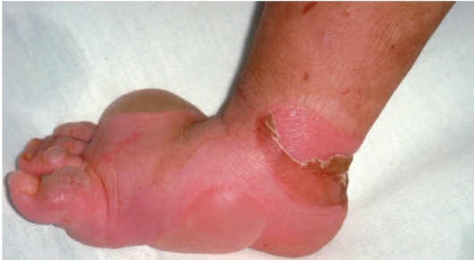
If the infection gets on the skin, the patient's condition may worsen. A typical change in the eyelids for ichthyosis - their eversion, is not observed. An additional symptom is whitish skin of the feet and palms with a thickening effect. If only these parts of the body are affected, another type of ichthyosis with different gene mutations is possible. To clarify the diagnosis, an external examination, anamnesis and the results of a histological examination are required.
This type of ichthyosis manifests itself in males in the first months of life. Most often, it looks like large skin scales in the form of plates of a black or dark brown hue. Sometimes such scales can lay on top of each other, creating a layer of keratinized skin in the form of a shell. Formations are observed on the back of the neck, scalp, buttocks and thighs, and are absent on the face, feet and palms. In some cases, corneal opacity is possible, therefore, in addition to a dermatologist, sometimes a comprehensive examination by specialized doctors, including an ophthalmologist, is required. 
A blood test may be required to confirm the diagnosis, as well as family genetic information and a general history. Relief of the disease can occur during the summer period of the year, while in a warm, humid climate. Frosty dry air can aggravate the disease. As a rule, no positive dynamics is observed with age.
Ichthyosis of the skin in children
If you find any suspicious formations on the skin in the form of scales or slight peeling in a child, you should contact a medical institution.
A similar hereditary disease can manifest itself during the development of the fetus in the womb at about the fourth or fifth month. At the same time, children are born with an altered skin structure - the presence of shells and scales of various sizes. The disease can affect the mouth, ears, eyelids, which can affect vision and food intake. With such a diagnosis, the appearance of membranes on the fingers and deformation of the skeleton are possible.
Most often, the disease manifests itself most actively before the age of three. The areas of peeling of the skin over time turn into keratinized skin with the so-called fish scales of a gray or dark shade, reddening of the skin with the presence of a film can be observed. The lines on the palms become more pronounced. The disease may be accompanied by itching, burning, painful sensations when the scales separate.
In addition to the external manifestation of the disease, the following changes are possible:
- Deterioration of the structure of hair and nails - their stratification and fragility.
- Conjunctivitis of the eye.
- Development of myopia.
- Allergic reactions.
- Kidney disease.
- Tooth decay and destruction of tooth enamel.
- Heart failure.
In such cases, you need to immediately start treatment on the advice of a doctor and under his supervision. For newborns, early therapy is provided - the child needs to maintain an optimal temperature and humidity, for which he is placed in a jug. A comprehensive examination of the patient is carried out by a surgeon, ophthalmologist and other specialized specialists. Further, additional treatment is prescribed. It will be chosen depending on the exact diagnosis - the type of ichthyosis and the severity of the disease.
When treating a child, it is important to take into account the psychological aspect, because an aesthetic change in the skin can affect his communication with peers.
Ichthyosis treatment
The outcome of the disease in most cases depends on the severity of the disease and the degree of gene mutation. With a mild degree of mutation and timely therapy, a successful treatment is possible, or at least an improvement in the patient's quality of life. A less favorable result is predicted in case of metabolic disorders and damage to other body systems, in addition to its genetic changes.
As a rule, treatment consists of moisturizing the skin - this is required to reduce dryness of the skin and reduce cracking. Prescribe medicinal ointments, creams, moisturizers based on lanolin or petroleum jelly, carry out additional keratolytic therapy. In some cases, baths with special solutions can be prescribed - soda, starch, salt, sometimes with the addition of chamomile or sage.
Of the drugs, vitamin complexes are sometimes prescribed - vitamins of the group are important for the skin A, E, WITH, V, if necessary - antibiotics, medicines to maintain the function of the thyroid gland, antifungal agents, iron preparations. It is important to maintain the body's immunity. So, in case of its weakening, immunomodulatory drugs can be recommended. In the course of treatment, it is required to regularly take urine and blood tests.
- prolonged exposure to the sun, overheating of the skin is not recommended;
- using soap can contribute to additional dryness of the skin;
- it is worth limiting the time spent in the cold - dry cold air can aggravate diseases;
- sanatorium treatment in places with a warm, mild climate will be favorable, in some cases swimming in the sea is allowed.
In any case, you cannot self-medicate, especially with such a serious diagnosis. A doctor's consultation and strict adherence to his recommendations can significantly improve the patient's quality of life and prevent the development of irreversible consequences.
When diagnosing ichthyosis, it is important to avoid hypothermia and prolonged exposure to frosty air, and also avoid overheating of the skin in the sun.
For any skin problems, you should consult a doctor, widespread peeling is no exception. An unpleasant diagnosis can be hidden behind it - ichthyosis. Due to its hereditary origin, treatment can be quite lengthy, difficult, and requires a lot of patience. It is important not to give up, because modern medicine offers many ways to make the patient's life easier and better.
Related Articles

Problems and solutions for cleaning reusable respirators Can the gas mask be washed
Machine for cleaning industrial premises with humidification
Ionizing radiation and protection against it Methods and methods of protection against ionizing radiation
Construction site information signs
Environmental problems of society at the present stage - file n1