Eye biomicroscopy is an objective method for studying the structures of the eye, which is carried out by a special device - a biomicroscope (slit lamp). Using this method, you can examine the elements of the anterior and posterior parts of the eyeball (learn about the eyeball).
Device structure
A biomicroscope consists of a lighting system, which is a light source, and a microscope for two eyes.
The light from the lamp passes through the slit-like diaphragm, after which it is projected onto the cornea or sclera in the form of an oblong rectangle. The resulting optical section is examined under a microscope. The doctor can move the light gap to those elements that need to be examined.
Indications and contraindications
In the pathology of what eye structures is biomicroscopy indicated?
- Conjunctivitis (conjunctivitis, education)
- Cornea (inflammation, dystrophic changes).
- Sclera.
- Iris (inflammation, structural abnormalities).
- The lens.
- Vitreous body.
Also, these methods are performed for cataracts, glaucoma, the presence of foreign bodies in the eye, at the stage of preparation for eye surgery and in the postoperative period.
There are no absolute contraindications to this diagnostic manipulation. The procedure should be rescheduled if the patient has an exacerbation of mental disorders or is in a state of intoxication.
Methodology
First, the preparation of the patient is carried out - drops are added to the eyes, expanding the pupil (if necessary, examination of the deep structures), or special dyes (in cases where it is necessary to diagnose corneal pathology).
The patient sets his head on a special stand with stops for the forehead and chin. The doctor is opposite the patient, moves the microscope and lamp to the eye level of the patient. Using the diaphragms, the size and shape of the light gap are regulated (more often in the form of a rectangle, less often in the form of a small circle). Rays of light are sent to the studied structures of the eye, after which they are examined in detail.

Examining the cornea, you can find foci of opacities, infiltrates, newly formed vessels. The biomicroscopy procedure allows you to clearly examine the lens, as well as to identify the localization of pathological changes. This method allows you to explore the blood vessels of the conjunctiva.
Also, using a biomicroscope, you can evaluate the sphericity and specularity of the cornea, determine its thickness, as well as the depth of the anterior chamber of the eyeball.
There are several lighting options during this diagnostic procedure:
- direct focused lighting - the light is directed to the studied area of \u200b\u200bthe eye. So evaluate the transparency of the optical media of the eyeball;
- indirect focused light - light rays are sent next to the studied area, as a result of which it is possible to better consider pathological changes due to the contrast of the illuminated and unlit area;
- reflected light - this is how certain structures (for example, the cornea) are examined by light reflected from other elements (the iris), like from a mirror.
Recently, ultrasound biomicroscopy of the eye has become increasingly popular, thanks to which it is possible to examine the lateral parts of the lens, the posterior surface and the cut of the iris, and the ciliary body.
Also find out how other examinations are performed by an ophthalmologist, for example, eye pressure measurements and is it scary? Read
For a more complete acquaintance with eye diseases and their treatment - use the convenient search on the site or ask a specialist.
) is a detailed study of the structures of the eye, carried out using a special optical device - a slit lamp. The main part of the device is a diaphragm in the form of a narrow slit, as a result of which it got its name.
The most common model of the slit lamp ShchL-56 in the Soviet Union. Using the lamp of this model, it is possible to examine both the anterior and posterior parts of the eye - the vitreous body and.
Biomicroscopy makes it possible to detect the smallest changes in the eye, detect small ones and determine the depth of the pathological process. Biomicroscopy is very important for the diagnosis of perforated wounds of the cornea and other eye diseases.
Biomicroscopy (a synonym for living eye microscopy) is a research method that allows you to examine in detail the conjunctiva, cornea, iris, anterior chamber of the eye, the lens, the vitreous, and also the central parts of the fundus (biomicrophthalmoscopy); proposed by Gulstrand (A. Gullstrand). The biomicroscopy method is based on the phenomenon of light contrast (Tyndall phenomenon).
With the help of biomicroscopy, you can conduct an early diagnosis of the majority (for example, glaucoma and trachoma), determine the perforated wound of the eyeball, detect very small foreign bodies in the conjunctiva, cornea, anterior chamber of the eye and the lens that are not detected by x-ray (glass, aluminum, coal, eyelash). Biomicroscopy is carried out using a slit lamp.
The device (Fig. 1) consists of a illuminator and a binocular stereoscopic microscope. The light source in the illuminator is a lamp (6 V, 25 W), powered by an 127 or 220 V AC mains through a step-down transformer. On the path of the light beam is
slot mechanism, allowing to obtain a vertical and horizontal lighting gap. In the case of the binocular microscope is an optical device that provides various magnification options (5, 10, 18, 35, 60 times). A scattering lens with a power of about 60 D is mounted on a binocular microscope, which neutralizes the positive effect of the optical system of the eye and allows you to see the fundus.
Fig. 1. Slit lamp ShchL-56: 1 - front installation; 2 - illuminator; 3 - binocular microscope; 4 - coordinate table; 5 - tool table.
Biomicroscopy is performed in a dark room, creating a sharp contrast between the darkened and lamp-lit areas of the eyeball. In the process of biomicroscopy, diffuse, direct focal light, indirect illumination (dark field), transmitted light, a moving beam, research in the illuminated zones (mirror field method) are used. The main type of lighting is direct focal. When focusing light on the cornea, an optical section of it is obtained in the form of a slightly opalescent convex-concave prism (Fig. 2). The front and back surfaces, the corneal substance itself, stand out well. If there is an inflammatory focus or clouding in the cornea, studying the optical section allows you to decide where the pathological focus is located, how deep the corneal tissue is affected; with a foreign body in the cornea - whether it is in the corneal tissue or partially promotes into the eye cavity, which allows the doctor to correctly determine the method of intervention.
When focusing light on the lens, an optical section of it is cut out in the form of a biconvex transparent body. In the section, the surfaces of the crystalline lens, as well as the grayish oval stripes, the so-called separation zones, due to the different density of the lens material, are clearly distinguished (Fig. 3). The study of the optical section of the lens allows you to see and accurately localize the beginning clouding of its substance, which is of great importance for the early diagnosis of cataracts. Focusing the light on the fundus allows one to examine the retina and optic nerve disc in the optical section (Fig. 4). This is important for the early diagnosis of optic neuritis, congestive nipple, centrally located ruptures of the retina.
Less diagnostic opportunities are opened up by biomicroscopy of translucent and opaque membranes of the eyeball, for example, conjunctiva, iris. However, in this case, biomicroscopy is an important addition to other methods of examining a patient with eye disease.
Fig. 2. Optical section of the cornea: a, b, e, d - the front surface of the cornea; 3, e - rib of the rear surface; b, d, d, e - the thickness of the cornea.
Fig. 3. Optical section of the lens: 1 - central gap; 2 - the central surface of the embryonic nucleus; 3 - peripheral surfaces of the embryonic nucleus; 4 - surface senile core; 5 - subcapsular cleavage zones; 6 - front and rear surfaces of the lens. Fig. 4. Optical section of the retina and optic disc.
26-07-2012, 20:39
Description
Research Methodology
It does not present any difficulty, since the main parts of the conjunctiva are well accessible for examination in a slit lamp. In some cases, when studying the conjunctiva of the eyelids, an assistant is needed, whose role is to twist and hold the eyelids.
With biomicroscopy of the conjunctiva of the eyelids, it is often necessary to examine carefully transition folds. When the upper eyelid is turned out, the upper transitional fold does not protrude forward enough and as a result cannot be examined with due care. To facilitate the study of transitional folds, V.P. Filatov in 1923 proposed introducing 2 ml of a 0.5% solution of novocaine under the conjunctiva. Transitional fold bulges forward. Novocaine spreads the folding of the conjunctiva, which makes the tissue more accessible to inspection. On the stretched conjunctiva, the follicles, papillae and scars that develop during trachoma are better visible.
Conjunctiva biomicroscopy can be used almost all lighting options. A general overview of the conjunctiva is usually performed under diffuse illumination under small magnifications of the microscope. Examination in an optical section with a narrow slit is advisable in the presence of edematous conjunctiva, follicular formations, conjunctival cysts.
Silhouettes of meibomian glands, cicatricial changes in the conjunctiva can be detected by indirect illumination.
- Diaphanoscopic illumination helps with differential diagnosis between translucent follicles and opaque papillary formations.
- The conjunctiva of the eyeball is a transparent, translucent tissue, so it can be examined in transmitted light. Rays of light penetrating freely through the conjunctiva fall on the sclera lying beneath it. A strongly lit screen forms, against which numerous conjunctival vessels, cystic formations are clearly visible.
- To study the condition of the vasculature of the conjunctiva, you can use the study in redless light (green filter). The vessels at the same time appear more distinctly in the form of dark branches on a blue and then green background.
- Examination by a sliding beam reveals various kinds of irregularities on the surface of the conjunctival tissue.
- When using the mirror field method, the conjunctival promising formations give a clearly visible peculiar intense brilliant reflex.
The conjunctiva is normal
Unchanged conjunctiva When examined with a slit lamp, the eyelid has a smooth, shiny surface without wrinkles and thickenings and appears translucent, which makes it possible to see its adenoid layer. The rich vascularization of the conjunctiva is noteworthy. The vessels of the conjunctiva of the eyelids are distinguished by the correct vertical arrangement of the main large branches. In diffuse light, perforating vascular branches arising from arterial arches located in the thickness of the upper and lower eyelids, posterior conjunctival vessels are visible. On the upper eyelid, three zones of these vessels can be distinguished (Fig. 14).
Fig. fourteen. Zones of the posterior conjunctival vessels of the upper eyelid. 1-first zone; 2-second zone; 3rd zone.
First zone represented by 8-10 rather short vascular trunks arising from the marginal arterial arch of the upper eyelid and appearing on the conjunctiva 2 mm from the free edge of the eyelid. Second vascular zone consists of a smaller number of longer perforating branches originating from the peripheral arterial arch of the upper eyelid and appearing on the conjunctiva, respectively, on the upper edge of the cartilage. Both vascular zones in the lower third of the conjunctiva of the upper eyelid anastomose with each other, forming in this place the third zone of connecting and interwoven vascular branches.
On the lower eyelid peripheral arterial arch often absent, and when examining the conjunctiva, only one area of \u200b\u200bthe posterior conjunctival vessels is visible, originating from the marginal arterial arch of the eyelid. Many small branches departing from the main arterial trunks, forming a more superficial vascular plexus.
The conjunctiva of the sclera is transparent and it is recognized mainly by the existing vessels. With biomicroscopy, it can be distinguished two vascular systems (fig. 15).

Fig. fifteen. Conjunctival vessels of the eyeball (optical section).
One of them, more superficial, subepithelial, consists of the posterior conjunctival vessels passing from the conjunctiva of the eyelid and anastomosing in the circumference of the limbus with the anterior conjunctival vessels. These vessels lie in the surface sections of the optical section of the conjunctiva. Another system of vessels is located more deeply, belongs to the category of episcleral. These vascular systems differ not only in the depth of location, but also in the color of the vascular trunks, their caliber, the possibility of displacement along with the conjunctiva of the eyeball during blinking movements of the eyelids.
Conjunctival superficial vessels they have a bright red color, are quite thin and branched, easily shift together with the conjunctiva when it glides along the surface of the eyeball.
Most of these vessels are usually visible granular blood flow - physiological phenomenon. It is not always possible to distinguish an artery from a vein in the direction of blood flow, since it changes from time to time. In some cases, especially with the expansion of blood vessels, oscillatory movements of the blood column in one direction or another are observed and complete stoppage of blood flow is a stasis phenomenon. When instilling vasoconstrictors, in particular adrenaline, the granular cree o-current is restored.
More deeply located vessels are more saturated in color, large caliber. When the conjunctiva is displaced, they do not change their location. Distinguish episcleral artery from a vein it is often quite difficult, since the difference in their color is hardly noticeable, and the direction of blood flow is almost impossible to determine due to the significant width of the vessels.
In the limb area The conjunctiva imperceptibly passes into the transparent tissue of the cornea. In many biomicroscopy, especially in the upper and lower limbs, this transition can be seen in the form of peculiar radial stripes of a whitish color: between these bands - conjunctiva cords - transparent sections of the cornea tissue are clearly visible. The alternation of transparent and opaque zones will give the limb a characteristic striation. This is the so-called zone of palisades (Fig. 16).

Fig. 16. The area of \u200b\u200bthe palisades.
Sometimes pigment is deposited about this area, due to which the radial striation appears more clearly.
In biomicroscopy, a limb region shows a very rich, peculiar architectonics, network of vessels, which are mainly branches of the anterior conjunctival arteries and veins. Here you can also highlight three zones of blood vessels (fig. 17).

Fig. 17. Limb vessels.
- The first, most peripherally located area of \u200b\u200bthe palisades is characterized by the presence of parallel, non-anastomosing vascular branches located about the corresponding radial depressions of the limb. Its length is 1 mm.
- Next, towards the cornea, there follows a second, middle zone, characterized by a large number of vessels anastomosing between themselves. Its length is 0.5 mm.
- The third - the zone of the final capillaries - occupies a space equal to 0.2 mm.
Normally, no matter how wide the limb, the end capillaries do not penetrate into the transparent tissue of the cornea. One of them does not end freely. At the apex of each vascular loop (end capillary), the direction of blood flow changes, becomes reverse, the vessel itself expands. This is the beginning of the venous knee of the capillary.
Limb vessel biomicroscopy plays a large role in the early diagnosis of trachoma.
When examining the limbal and perilimbal regions, one can see vessels containing very light (diluted) blood, and sometimes a colorless liquid. it water veinsdescribed in 1912 by Ascher. Histologically found that they depart from the scleral wall of the Schlemm's canal, perforate the sclera in an oblique direction, and appear on its outer surface in the circumference of the limb.
Water veins visible in every third to fourth patient, mainly in the region of the palpebral fissure, slightly above or below the horizontal meridian of the eye. The number of visible veins is individually different. If it is not always possible to immediately notice a vein, then the conjunctival or episcleral vessel that perceives it is usually well visible. In some of these vessels, it is possible to see two fractions of a liquid that are different in color (blood and clear, aqueous humor). The vessel in these cases appears double layer, and sometimes three-layer (Fig. 18).

Fig. eighteen. Water vein.
When the endothelial septa disappear between these layers, the fluids stick together into one common current and the vessel (vein) takes on a light pink color, and then a normal red color. If you trace the progress of such a vessel to the limb, then you can see a water vein.
During prolonged observation of the place where the water vein flows into the receptive vessel, Z. A. Kaminskaya (1950) saw the phenomenon she called piston phenomenon. From time to time, often synchronously with the pulse, a small column of blood is poured into the water vein and then casts back. This phenomenon resembles a pump, which, as it were, pumps out the intraocular fluid inside it from a water vein. According to Z. L. Kaminsky, the piston phenomenon plays a role in the mechanism of intraocular fluid removal along the anterior outflow path.
With conjunctival biomicroscopy, especially with glaucoma, attention should be paid anterior ciliary vesselsassociated with sclera emissaries. They are visible at some distance from the limb. Arteries enter the eye, veins exit it.
It is difficult to distinguish an artery from a vein even with a slit lamp. The artery is usually convoluted stronger than a vein, and has fewer lateral branches. For a more accurate differentiation of an artery from a vein, you need a food thread vessel (after instilling anesthetics) with the edge of a glass rod. If the central segment of the vessel expands and overflows with blood, then this is a vein, if it is a peripheral segment, then the vessel is an artery.
With age conjunctiva is subject to change. In older people, there is a thinning of the optical section of the conjunctiva, a decrease in the transparency of the tissue, which acquires a yellowish tint. In the area of \u200b\u200bthe palpebral fissure in the conjunctiva of the eyeball, fatty and hyaline deposits are often observed. The conjunctival and episcleral vessels become denser, become crimped. An examination in the optical section shows that they raise the conjunctival tissue above themselves, protruding above its surface. Often there is varicose veins with the formation of petechiae.
Pathological conjunctival changes
Conjunctival diseases occupy one of the leading places among other types of ocular pathology, making up, according to various authors, from 30 to 47% of the total number of eye diseases.
Inflammatory diseases
The conjunctiva is widely in contact with the external environment and is therefore most susceptible to inflammatory diseases associated with the introduction of an exogenous infection.
Trachoma
Trachoma- chronic infectious proliferative inflammation of the conjunctiva, characterized by tissue hypertrophy with the development of follicles and papillae in it and ending with scarring.
Trachoma belongs to the group of those diseases in which biomicroscopy is leading research method throughout the clinical course of the process. Microscopy is necessary for the early diagnosis of trachoma, determining its stage, monitoring the dynamics of the disease under the influence of tone or other therapy, which allows you to determine when it is necessary to strengthen, weaken or change the treatment. A large role belongs to biomicroscopy in determining the cure of the patient. Dynamic monitoring of patients with trachoma shows that in most cases only a biomicroscopic examination allows you to establish the true recovery of the patient, the complete elimination of the trachomatous process.
Clinical manifestations of trachoma varied - from pronounced to subtle changes in the conjunctiva and limb. There are worn and light forms of trachoma. In the latter case, biomicroscopic examination is extremely important for reasons of an epidemiological nature.
When examining a patient with the naked eye in the early, initial period of the trachoma, the conjunctiva in some cases may seem almost unchanged. Researchers are attracted only red dots on the conjunctiva of cartilage.
- In biomicroscopy, these points appear to be expanded, newly formed capillaries extending from the main vascular trunks of the conjunctiva and their branches in the direction perpendicular to the conjunctival surface. With the development of the process, each of these vessels begins to branch, forming capillary arches (vascular bouquets) located parallel to the surface of the connective membrane.
- An examination in the optical section shows that the vessels lie under the epithelium in the adenoid tissue of the conjunctiva. In the circumference of each vascular stem, the conjunctival papilla is formed. Group clusters of hypertrophied papillae are more often visible on the conjunctiva of the cartilage of the upper eyelid, mainly in the region of the corners of the eyelids. where in connection with this a peculiar picture of the mosaic arises.
However, early biomicroscopic diagnosis of trachoma, based only on a statement of an increase in the number and hypertrophy of the papillary conjunctival formations, may be erroneous. Papillary hypertrophy observed with a number of banal chronic conjunctivitis with a benign course and a favorable outcome.
Dynamic monitoring of patients with trachoma soon after the detection of hypertrophy and an increase in the number of papillae, and sometimes in parallel with them, reveals the presence of follicles. They appear on the conjunctiva of the transitional fold, and then the cartilage, located in the diffusely infiltrated tissue between the papillae, as if pushing and squeezing them to the sides (Fig. 19).
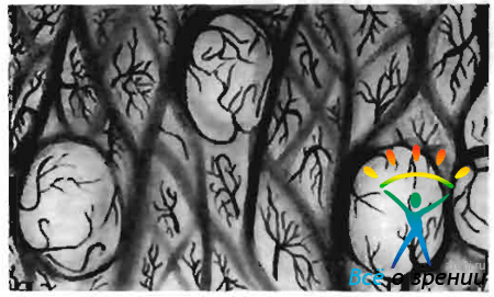
Fig. 19. Stage I trachoma. Changes in the conjunctiva of the century.
Follicles, unlike papillae, develop not only in the conjunctiva of the eyelids, but also on the lacrimal flesh and lunate fold.
Initial follicles they have the appearance of gray, slightly protruding above the surface of the conjunctiva, mildly contoured formations located mainly in the places of vascular bifurcation; they still do not have their own vessels. As each follicle grows and ripens, newly formed vessels are sent to it from the surrounding tissue, which braid it like a net, giving at the same time branches that penetrate into the depths of its tissue.
In some cases distinguishing papillae from follicles is not easy. An inexperienced researcher can take the papillae for the follicles and vice versa. For the purpose of correctly recognizing them during examination with a slit lamp and the correct interpretation of the process, it is recommended to dry the surface of the conjunctiva with a moist sterile swab before examination, while removing any mucus deposits and tears. When differentiating the papilla from the follicle, the appearance of the formation, its size, shape, color, degree of transparency, and the nature of vascularization are taken into account.
The conjunctival papilla has a smaller size compared to the follicle, a polygonal shape, a more saturated red color. Its fabric is only relatively transparent. The character of the vascularization of the papilla is typical. The feeding vessel is located inside it (in the center or slightly eccentric, Fig. 20),
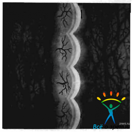
Fig. twenty. Stage I trachoma. Papillae of the conjunctiva of the eyelid (optical section).
to the appearance of the vessel, as a rule, precedes the formation of the papilla.
Trachomatous follicle the papilla is 4-6 times larger, has a spherical shape, gray-yellow color. Its tissue is more transparent than the tissue of the papilla. The follicle has a sharply different type of vascularization from the papilla. The vessels are located mainly on the surface of the follicle (Fig. 21)

Fig. 21. Stage I trachoma. Conjunctival follicles of the eyelid (optical section).
and develop later than the follicle itself.
In I steel trachomaIn addition to follicles and papillae, biomicroscopic examination reveals a change in the epithelium and diffuse cellular infiltration of adenoid subepithelial tissue. The layers of the epithelium are thickened, less transparent than normal. Adenoid tissue is swollen, loose, granular, which makes the optical section of the conjunctiva significantly thicker and less transparent. In the vessels of the conjunctiva stagnation of blood with the presence of small hemorrhages in the circle. The correct course of the vessels is violated, numerous anastomoses appear between them.
In stage II trachoma many papillae undergo reverse development. Only with the papillary form of trachoma is a well-developed mosaic of papillae visible throughout the conjunctiva of cartilage. There is an increase in the number of follicles, but along with this, some of them undergo collicative necrosis in the center. Such follicles acquire a dull gray color, fuzzy borders, often open. The scarring process begins.
The identification of initial, even hidden, deeply located scars helps maximum narrowing of the lighting gap and maximum brightness of the light beam during biomicroscopy. Scars that occur at the site of the follicles look like very delicate white lines located between the papillae. They must be distinguished from between the papillary fissures, usually made by mucus and leukocytes.
In the circumference of the scars there is a significant number of newly formed vessels (Fig. 22).

Fig. 22. Scars of the conjunctiva of the eyelid with trachoma.
In stage III trachoma is characterized by progressive scarring, leading to connective tissue replacement of the affected conjunctiva. During biomicroscopy, islands of infiltration and hypertrophied papillae are visible among smooth, shiny, well-defined scars.
With stage IV trachoma silver cicatricial strands are found, located mainly in places of richer vascularization of the conjunctiva. Marked scarring of the conjunctival tissue is noted in the region of sulcus subtarsalis, i.e., there. where the main trunks of the posterior conjunctival vessels arise, as well as in the area of \u200b\u200banastomoses between the individual vascular branches. Scars are usually located along the vessels or intersect them at an angle. In the latter case, against the background of vascular trunks, the scars are more prominent.
In the end untreated or poorly treated trachoma scar tissue completely kneads all follicles, papillae and leads to obliteration of blood vessels. An optical section of the conjunctiva in the area of \u200b\u200bthe formed scar in these cases cannot be obtained.
In the end successfully treated trachoma scars also develop, however, they are tender, translucent, do not constrict the conjunctival tissue, and do not lead to the closure of the excretory ducts of the conjunctival glands. Against the background of such scars, the section of the conjunctiva seems almost normal. Scars can be seen only on the delicate silvery layers of a denser tissue, visible at different depths of the optical section.
Biomicroscopic studies have shown that with trachoma, in parallel with changes in the conjunctiva, and sometimes preceding it, they develop changes in the limb. There is superficial, diffuse vascular keratitis, or pannus.
It allowed to come to the correct understanding of the trachomatous process of the cornea and to regard it not as a complication, but as one of the components of the sometimes early clinical manifestation of trachoma. It is proved that in some cases the cornea can be the site of the primary localization of the trachomatous virus.
According to the observation of a number of authors (L. S. Slutskin, 1940; N. N. Nurmamedov, 1960), damage to the cornea during the trachomatous process in the study slit lamp observed in 95-100% of patients. When examined by conventional methods with the naked eye, pannus is detected only in 7-10% of patients (V.V. Chirkovsky, 1953).
When viewed with a slit lamp, it can be seen that already early trachoma the transparency of the upper limb decreases, the characteristic radial striation disappears. The limb takes on a grayish tint and protrudes slightly. surface of the cornea, its border becomes uneven. The vessels of the limbus are usually overflowing with blood and are visible to the smallest branches.
Soon, when examining in transmitted light in the upper limb, it is possible to notice very gentle cloudy clouding of the corneaconsisting of a mass of gray dots and thin threads. In the optical section, the corneal tissue in this zone appears to be opalescent; opacities are located in the subepithelial zone. The number of limbal vessels increases, from which capillaries extend, penetrating the corneal tissue along the grayish filaments-infiltrates. These vessels, like infiltrates, are very superficial (Fig. 23).

Fig. 23. Trachomatous pannus (optical section)
Pannus vessels consist mainly of winding, densely branching veins; the artery has a more rectilinear course and are located deeper.
A little later in the limb zone you can see small, round, grayish gelatinous islets - follicles. They, like conjunctival follicles, go through the whole cycle of their development; in some cases they have an abortive development. Often the follicles fused together form a zone of pronounced infiltration, visible to the naked eye. In the end, small cicatricial impressions covered with epithelium remain in place of the follicles. These impressions are facets, also known as “ocelli”, arise as a result of degeneration and decay of trachomatous follicles.
In the malignant course of the pannus, its ulceration, infiltration and blood vessels penetrate into the deeper layers of the cornea. In such cases, they are visible in the middle and deep parts of the optical section. In this case, secondary changes may also develop - deposition in the affected cornea of \u200b\u200bcalcium, lipoids. The outcome of the trachomatous pannus remains more or less intense, with a tendon hue, clouding of the cornea. Penetrated by vessels.
When making a diagnosis of trachoma, one must take into account the possibility of mixing this disease with other inflammatory lesions of the conjunctiva.
In the differential diagnosis of trachoma with follicular conjunctivitis, it should be borne in mind that with follicular conjunctivitis absent diffuse infiltration of subepithelial tissue and hypertrophic papillae. When examined with a slit lamp, the follicles appear small, transparent, without a developed network of capillaries (Fig. 24).

Fig. 24. Follicular conjunctivitis.
With follicular conjunctivitis, as well as with acute conjunctivitis of a different origin, thickening of the limb, expansion of the terminal capillaries and a slight lengthening of individual capillary loops can be observed. However, these changes soon disappear. Biomicroscopic examination does not detect any newly formed vessels or infiltration in the corneal tissue. Pannus with follicular conjunctivitis does not happen; there is no scarring of the conjunctiva in the outcome of the disease. By eliminating the process, the tissue of the conjunctiva appears completely unchanged.
In the differential diagnosis of spring catarrh trachoma the pale, sometimes milky white color of the conjunctiva, the predominant localization of changes in the conjunctiva of the cartilage of the upper eyelid (tarsal form) are taken into account. A lethal examination of these changes by the method of binomnoscopy can be performed only after removing a thick coating of viscous secretion from their surface.
Macroscopically detected conjunctival surface roughness. This is due to the presence of dense, pale, shiny, as if polished, formations. These growths are known to be based on hyaline degeneration of subepithelial tissue.
Hyaline growths on the conjunctiva with spring catarrh, they are fused, degeneratively degenerated papillae. When examined with a slit lamp, they look like multiple flat, polygonal and oval, adjacent to each other formations with a smooth surface. Visible vascular bundles extending from large arterial trunks and heading perpendicular to the surface of the conjunctiva. From these bundles, a more superficial network of vessels arises (Fig. 25, a).

Fig. 25.Spring Qatar. a - tarsal form: b - limbal form.
Unlike papillae in the spring of Qatar papillae with trachoma smaller, red, more cloudy and lack luster due to infiltration and desquamation of the epithelium. In addition, the papillae during trachoma never merge into one continuous mass. Each is formed around one vascular stem and is separated from neighboring formations by a more or less narrow gap.
In some cases, you have to differentiate trachomatous pannus with limbal form of spring catarrh. With a cheerful catarrh, changes occur around the entire limb, and not only in its upper half, as with trachoma. A limb forms small grayish islands, consisting of a hyaline glassy substance that is poorly translucent when examined in direct focal light. Often they merge and form a continuous roller with a tuberous surface, sometimes approaching the cornea (Fig. 25, b). Vascularization of the vitreous formations compared with vascularization with trachomatous pannus is very small. The vessels of the limbus usually pass through them at various depths and singly end on the cornea.
In the spring of Qatar damage to the more centrally located parts of the cornea is also observed. Here, in the most superficial layers, very small, flat scaly overlays of gray-white color appear. When the eyelids blink, these scales can be washed off by a tear and erosion remains in their place. It is believed that these are neurotically degenerated elements of the corneal epithelium. Their favorite location is the upper half of the cornea (under the upper eyelid) and the region of the palpebral fissure.
It should not be forgotten that with the limbal form of spring catarrh cartilage conjunctiva is often involved in the process. It acquires a milky white hue, against the background of which, even under a small magnification of the microscope, a typical mosaic of papillae is observed. They also noted hyaline tissue degeneration, which gives the papillae a peculiar, glassy, \u200b\u200btranslucent appearance.
At the end of spring Qatar all symptoms disappear without a trace; unlike trachoma, no scars remain. Changes in the limbus and cornea also undergo a complete reverse development. As a result, retrospective diagnosis of spring catarrh with a slit lamp is very difficult.
Dystrophic changes
Pingvekula- hyaline degeneration of the conjunctiva. It has the appearance of an islet of yellow color, most often located at the inner limb of the cornea. Biomicroscopic examination in direct focal light reveals that the process of degeneration captures the deep sections of the optical section of the conjunctiva. Vitreous forks amorphous masses are also detected under the conjunctiva. Sometimes small cavities are visible in the pingvecule tissue (Fig. 26).
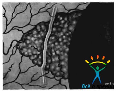
Fig. 26. Pingvekula.
Pterygium, or pterygoid, - a fairly common dystrophic change in the conjunctiva. When examining a slit lamp, special attention should be paid to examining the head of the pterygium, i.e., that part of it that is located on the cornea.
Two zones are distinguished in the head of the pterygium: vascular and non-vascular (Fig. 27).

Fig. 27. Pterygium.
The latter is located in front of the vascular zone (towards the center of the cornea) and consists of foci of turbidity of a gelatinous appearance, the processes of which extend to the deeper sections of the corneal stroma.
Biomicroscopic examination allows you to determine whether the pterygium is stationary or progressive, which helps to correctly resolve the issue of the timing and type of surgery. Stationary pterygium is characterized by a mild, flat, avascular zone of the head, which quietly merges with the tissue of the cornea. With progressive pterygium, the avascular zone is more pronounced, loosened and significantly rises above the surface of the cornea. In front of the head of the pterygium, there are sometimes point subepithelial opacities.
The differential diagnosis of pterygium with the initial form of Boven's epithelium - see below.
Neoplasms
Papilloma- a benign fibroepithelial formation, most often localized on the conjunctiva of the eyelids in the circumference of the lacrimal meat, less often on the conjunctiva of the eyeball. The tumor has a pink color, soft consistency, loosely soldered to the underlying tissues, often sits on a leg.
With biomicroscopy, it is possible to identify that the surface of the neoplasm is uneven, represents a conglomerate of papillomatous growths of a mosaic nature. His kind the tumor resembles a mulberry or cauliflower (fig. 28).

Fig. 28. Papilloma of the conjunctiva.
The tissue of the papilloma does not pass the light of the slit lamp, which makes it impossible to obtain an optical slice during bnomnoscopy. Papilloma is characterized by rather scarce vascularization, it lacks glomerular vascular formations typical of malignant and vascular tumors.
With the localization of papilloma in the limb, it is necessary to conduct differential diagnosis with carcinoma.
Epithelioma, or carcinoma, - malignant epithelial tumor of the conjunctiva; prone to active growth and relapse after removal. The tumor is localized, as a rule, in the circumference of the limbus. In the initial stage, it is difficult to distinguish from papillomas, pingvecules and the beginning pterygium.
With biomicroscopic examination a hallmark of epithelium is tuberosity, lobation of the tumor. Often, ulceration of the entire surface is observed, which is determined by the presence of a defect in the optical section of the conjunctiva. This symptom is especially well detected after staining the surface of the epithelium with a fluorescein solution. In the differential diagnosis of epithelium, great importance should be given to the study using a slit lamp. nature of vascularization.
Epithelioma is richly vascularized. Each lobule of the tumor is equipped with a central vessel with a mass of capillary branches. The vessel penetrating the lobule of the tumor rises to its apex, and then descends back (Fig. 29).

Fig. 29. Conjunctival epithelium.
The basal capillaries form a common vasculature, which with its numerous anastomoses nourishes all segments of the tumor. This type of vascularization is pathognomonic for epithelium.
Epithelial intraepithelial localization, without violating the basement membrane of the epithelial layer, stands out as a separate form of precancerous diskeratosis - Boveia epithelium. The formation has the form of a grayish-white flat plaque, localized, as a rule, in the circumferential) ”limbus. When examined with a slit lamp, a roughness of the neoplasm surface is noted, which is revealed but a break in the light gap, a significant amount of white flakes of desquamating epithelium (diskeratosis). The boundaries of the tumor are clear. With Boveia epithelioma, a pronounced vascular reaction from the surrounding conjunctiva is observed
Nevus, or birthmarkconjunctiva does not belong to the category of true tumors. It is characterized by slow growth during the development of the body, in adults it becomes stationary. However, in some cases, malignant degeneration of the non-tissue is possible as a result of irritation, trauma to the nevus, and sometimes for no apparent reason.
Nevus is most often localized on the conjunctiva sclera in the limb. It can manifest itself in a pigmented and non-pigmented form, which leads to a different color of irreplaceable spots, from dark brown to light yellow.
Biomicroscopy in direct focal light and with a sliding beam reveals flat or very slightly prominent formation on the surface of the sclera, with fairly clear boundaries (Fig. 30).

Fig. thirty. Nevus conjunctiva.
Gentle, dusty pigmentation of the tissue is characteristic. With a narrow gap, you can determine the depth of the pigment, find out whether the nevus is epithelial or subepithelial. With subepithelial localization, pigment islands are visible in the optical section under the epithelial band. Sometimes there is a nevus with vacuum degeneration - the so-called naeviis cysticus. In the optical section with this form of nevus, numerous transparent cavities are visible - vacuoles separated by thin partitions.
There are vessels in the irreplaceable tissue, but they are few in number and do not give a dense branching.
Thorough biomicroscopy makes it possible to carry out a differential diagnosis between nevus and incipient conjunctival melanoblastoma. A large role in this belongs to the recognition of changes in the nature of vascularization. Detection of new vascular branches in the irreparable tissue is suspicious of its malignant transformation. Simultaneously with a change in the nature of vascularization, or somewhat later, an increase in the size of the neoplasm, increased pigmentation of the tissue, redistribution of the pigment is noted.
Conjunctival melanoblastoma refers to the most malignant tumors, has a tendency to metastasis. She. arises spontaneously or develops from the nevus of the conjunctiva. The tumor is localized most often around the limb, but is also observed in the lacrimal flesh, semilunar fold of the conjunctiva. Melanoblastoma spreads quite quickly, giving daughter growth nodes.
With biomicroscopy, the lobular structure of the tumor tissue, its enhanced pigmentation, is noted. Unlike nevus, the melanoblastoma pigment is coarser, lumpier. When trying to obtain an optical section in the region of melanoblastoma, the density of the tumor tissue, its intimate adhesion to the underlying scleron, is noted. A ray of light in the mass of the tumor usually penetrates weakly. A characteristic biomicroscopic feature of melanoblastoma is its magnificent vascularization, which is not observed with nevus. In the center of each lobule of the tumor, a branching bundle of capillary vessels is visible. In addition, there is a rich vascular network deep in the neoplasm.
We present one of our clinical observations, where the study of a slit lamp greatly helped to make the correct diagnosis.
Patient S., 30 years old. entered the hospital about neoplasms of the conjunctiva of the right eye. Since childhood, marks a pigmented spot on the eyeball, in recent years it has begun to increase in size and withstand above the surface of the eye.
When viewed in the upper parts of the limb unevenly pigmented conjunctival formation of elastic consistency, rounded 4X6 mm in size. The remaining parts of the eyeball are not changed. Visual acuity 1.0.
When examining with a slit lamp in direct focal light, it is clearly seen that the neoplasm has a tuberous surface. It is not possible to obtain an optical cut of tissue, which emphasizes its density. Marked vascularization of the neoplasm is noted (a large number of newly formed vascular loops). In the tumor tissue there is a lot of coarse lumpy pigment (Fig. 31, a).

Fig. 31. Conjunctival melanoblastoma. a - biomicroscopic picture; b - histological picture (stained with hematoxylin-tosine. magnification 10 X 20).
Diagnosis: conjunctival melanoblastoma, developed from nevus. Biomicroscopy in fluorescent lighting and a study with radioactive phosphorus, which showed a high level of accumulation of P confirmed the diagnosis.
Produced removal of melanoblastoma with preliminary and sequential diathermocoagulation of surrounding tissues. Histological examination revealed melanoblastoma of the alveolar structure with magnificent growth in the central departments. Cellular and nuclear atypisis of tumor tissue (epithelioid type of structure) were noted (Fig. 31. b).
Glaucoma Changes
Biomicroscopy of the conjunctiva of the eyeball Mandatory for a comprehensive examination of a patient with suspected glaucoma, as well as in the process of dynamic monitoring of patients with glaucoma. In both cases, it is necessary to pay attention to the condition of the anterior ciliary vessels (which play an important role in the outflow of intraocular fluid), especially those located in the upper and lower parts of the eyeball. Changing the vessels located within the palpebral fissure in frequently exposed to adverse environmental influences can disorient the observer. These vessels, especially in the elderly, are often tortuous, their varicose branches.
In glaucoma, there is a change in both the anterior ciliary arteries and the anterior ciliary veins; the latter with congestive glaucoma are changed more often. Particular attention in biomicroscopy should be given scleral foramen - emissaries through which the anterior ciliary arteries enter the eye and the veins exit. In patients with glaucoma, it is sometimes necessary to observe peculiar changes, called the emissary symptom.
Exist incomplete and complete symptom of an emissary, the first is more common than the second. An incomplete symptom of an emissary is expressed in an increase in the size of the scleral foramen by 2–3 times. With bnomnoscopy, it has the appearance of a grayish rounded spot, in the center of which (in some cases, eccentric) is the anterior ciliary vessel. Sometimes, near the expanded emissary, there are gentle accumulations of pigment brought here by flowing chamber moisture.
With a complete symptom of an emissary elevation, swelling of the conjunctiva occurs above the enlarged scleral foramen (Fig. 32),

Fig. 32. The complete symptom of emissary in glaucoma.
similar to that observed after fistulizing antiglaucomatous surgery. In some cases, such a conjunctival “cushion” does not appear above the emissary itself, but somewhat deviates from it. Its development is associated with the detachment of the conjunctiva from the sclera flowing through the emissary by intraocular fluid. A study in direct focal light reveals a layer of clear fluid under the conjunctiva. When the anterior ciliary vessel is located near the limb, the appearance of an emissary symptom is usually not accompanied by the formation of a typical conjunctival pad, since the latter in the limb region is rather tightly soldered to the underlying sclera. Under these conditions, as a rule, there is only a barely noticeable elevating conjunctival ridge.
Emissary symptom detection with biomicroscopy, the doctor is obliged to suspect the presence of a glaucomatous process. If during the examination of the patient, intraocular pressure is normal, then in order to detect glaucoma, special tests should be performed. Under load conditions, compensation due to the expansion of the emissary is often insufficient, which is reflected in an increase in intraocular pressure.
In relatively young patients, when the sclera is still not so dense, an emissary symptom often occurs in the early period of glaucoma or in preglaucomatous state. In elderly patients, due to the compaction of the outer shell of the eye, an emissary symptom appears in the later stages of glaucoma against the background of dystrophic changes in the sclera. The sclera around the anterior ciliary vessel is sometimes reduced and thinned to such an extent that the choroid becomes visible in the hole.
In the diagnosis of glaucoma, a certain value may have water vein monitoring. The early signs of glaucoma, according to Z. A. Kaminsky, include the appearance of a negative ebb phenomenon. When a vessel receiving a water vein is squeezed with a glass rod, a twofold reaction can be observed: either the vein remains transparent and the intraocular fluid fills the receptor vein (a positive ebb phenomenon), or the vein is filled with blood (a negative ebb phenomenon).
In addition to the negative phenomenon of reflux, glaucoma is peculiar plus-minus phenomenon. It consists in the fact that after squeezing a vessel receiving a water vein, the vein initially remains transparent, and then it is filled with blood. With glaucoma in a state of decompensation, all water veins are filled with blood, the piston phenomenon is absent.
It must be performed by patients who have undergone a fistulizing antiglaucomatous operation. After trepanation of the sclera by Elliot, anterior sclerectomy, an operation of iridencleosis over the upper limb, a filtrational, sometimes multi-chamber cushion is formed. An examination in direct focal light shows that its cavities are filled with a clear intraocular fluid. Through them, you can consider the filtering operating hole in the sclera, a block of pigment.
- With a well-defined filtering postoperative scar, normal numbers of intraocular pressure are usually observed.
- In case of unsatisfactory filtration, the surgical scar appears flat, abundantly vascularized due to the newly formed conjunctival and episcleral vessels.
Article from the book:.
Biomicroscopy Slit lamp inspectionDeveloper: Medelit Studio, KSMU 2006
Biomicroscopy - This is an intravital microscopy of the eye tissue, a method that allows you to examine the front and rear sections of the eyeball under various lighting and image sizes.
The study is carried out with using a special device - a slit lamp, which is a combination of a lighting system and a binocular microscope (Fig. 1).

Fig. 1. Biomicroscopy using a slit lamp.
Through the use of a slit lamp, you can see the details of the structure of tissues in the living eye.
The lighting system includes a slit-shaped diaphragm, the width of which can be adjusted, and filters of various colors. A beam of light passing through the slit forms a light section of the optical structures of the eyeball, which is examined through a slit lamp microscope. By moving the light gap, the doctor examines all the structures of the anterior part of the eye.
Patient's headset on a special stand slit lamp with emphasis on the chin and forehead. In this case, the illuminator and the microscope are moved to the eye level of the patient.
The light gap is alternately focused on that tissue eyeballto be inspected. The light beam directed to the translucent fabrics narrows and increases the luminous intensity to obtain a thin light section.
In the optical section of the cornea, you can see foci of clouding, newly formed vessels, infiltrates, assess the depth of their occurrence, identify various tiny deposits on its posterior surface. When examining the marginal loop of the vasculature and the vessels of the conjunctiva, one can observe the blood flow in them, the movement of blood cells.
With biomicroscopy it is possible to clearly consider the various zones of the lens (anterior and posterior poles, cortical substance, nucleus), and in case of violation of its transparency, determine the localization of pathological changes.
Anterior layers of the vitreous body are visible behind the lens.
Distinguish four biomicroscopy methods depending on the nature of the lighting:
- in direct focus lightwhen the light beam of the slit lamp is focused on the studied area of \u200b\u200bthe eyeball. In this case, it is possible to evaluate the degree of transparency of optical media and identify areas of turbidity;
- in reflected light. So you can consider the cornea in the rays reflected from the iris, when searching for foreign bodies or identifying areas of swelling;
- in indirectly focused lightwhen the light beam is focused next to the studied area, which allows you to better see the changes, thanks to the contract of highly and dimly lit areas;
- with indirect diaphanoscopic transmissionwhen reflecting (mirror) zones are formed at the interface of optical media with different refractive indices of light, which allows one to study tissue sections near the exit point of the reflected light beam (study of the angle of the anterior chamber).
With the specified types of lighting you can also use two methods:
- conduct research in a rolling beam (when the handle of the slit lamp moves the light strip along the surface from left to right), which allows you to detect uneven terrain (corneal defects, newly formed vessels, infiltrates) and determine the depth of these changes;
- perform research in a specular field, which also helps to study the surface topography and still reveal irregularities and roughness.
Use for biomicroscopyin addition, aspherical lenses (such as the Gruby lens) make it possible to perform ophthalmoscopy of the fundus (against the background of drug mydriasis), revealing subtle changes in the vitreous, retina and choroid.
The modern design and fixtures of slit lamps also make it possible to additionally determine the thickness of the cornea and its external parameters, evaluate its specularity and sphericity, and also measure the depth of the anterior chamber of the eyeball.
(Greek, bios life + mikros small + skopeo observe, investigate) is a special research method that makes it possible to examine in detail optical refractive media and tissues of the eyeball.
B. G. was first proposed by A. Gullstrand in 1911. The method is based on the phenomenon of light contrast (Tyndall phenomenon).
With B.'s help, it is possible to detect the smallest changes in the eye caused by a disease or injury, to diagnose very small foreign bodies. The method is of great value in the diagnosis of a number of eye diseases (e.g. trachoma, glaucoma, cataracts, neoplasms of the organ of vision, etc.).
The study is performed using a special device - a slit lamp (see). The domestic slit lamp SHL-56 combines a powerful illuminator (500 thousand lux) and a binocular stereoscopic microscope with a resolution of X5 to X60. The microscope is placed directly in front of the test tissue, the illuminator is on the side. The angle between them is called the angle of biomicroscopy. It varies within + 60 °. The study is conducted in a dark room. The sharp contrast of the darkened and illuminated areas of the eye allows you to see details that are indistinguishable under normal lighting.
In the process of B. g., The following lighting methods are used: direct focal, parafocal, oscillatory, transmitted light, moving beam, mirror field. Using special devices, inspection can be done in the infrared and ultraviolet rays of the spectrum, luminescent, polarized light.
Fig. one. Optical section of the cornea: a, b, c, d - the front surface; d, e, f, z - the back surface; b - e and g - s - the thickness of the cornea. Fig. 2. Türk line in biomicroscopy (whitish dots): on the left - g in transmitted light; on the right - in the optical section of the cornea.
A study in direct focal illumination allows one to obtain an optical section (optical section) of the cornea, lens, vitreous body, retina and optic disc. The optical section of the cornea has the form of a slightly grayish, opalescent prism (Fig. 1), the width of the cut depends on the width of the transmitted light beam. Normally, the incision is dotted with gray dots and strokes - this is how fibrils and nerves of the cornea dissected by a beam of light look like. If there is an inflammatory focus or clouding in the cornea, the optical incision makes it possible to solve the question of exactly where the pathological focus is located, how deeply the corneal tissue is affected. If there is a foreign body, an examination in the optical section helps to establish where it is located - in the cornea or penetrates into the cavity of the eye, which correctly guides the doctor in choosing the method of intervention.
At B., the Türk line is easily revealed, edge meets in 50% of cases when studying healthy eyes, mainly in children. The Türk line is unstable, its formation and characteristic location are associated with the thermal current of the intraocular fluid. The cooling of the fluid moving along the posterior surface of the cornea, and the consequent slowing of its current velocity, leads to the deposition of cellular elements suspended in chamber moisture on the cornea. The line is located on the posterior surface of the cornea, vertically below, and reaches the level of the lower pupillary edge. It consists of white blood cells and lymphocytes, the number of which varies from 10 to 30. In transmitted light, the cellular elements look like translucent deposits, in direct focal light they take the form of whitish dots (Fig. 2).

When focusing light and a microscope on the lens (direct focal light), an optical section of the lens in the form of a biconvex transparent body is cut out (see. Lens). The section shows grayish oval stripes - the separation zone, due to different densities of the lens material (Fig. 3). The inner surfaces of the embryonic nucleus (1) with embryonic sutures, indicated by black Y-shaped lines in the figure, the outer surface of the embryonic nucleus (2), the surface of the senile nucleus (3), cortical substance (4), cleavage zones (5), the front and back surface of a crystalline lens (6). The study of the optical section of the lens makes it possible to see and accurately localize the gentle initial turbidity of its substance, which is of great importance in the early diagnosis of various kinds of cataracts.
Using the method of biomicroscopy of the vitreous body, gray fibrillar structures (the skeleton of the vitreous body) that are indistinguishable by other methods are revealed in it. The study of these structures has a certain diagnostic value, especially with myopia.
Fundus biomicroscopy (bio-microphthalmoscopy), fundus fundus biomicroscopy in the spectrum (biomicrochromophthalmoscopy) open up new possibilities in ophthalmoscopic diagnosis (see Ophthalmoscopy). The use of direct focal light allows you to see the optical section of the retina and optic disc. The retina is detected in the form of a concave-convex translucent grayish strip located between the vitreous body and the choroid itself. The study of the optical cross section of the retina helps to diagnose and accurately localize small hemorrhages, vascular microaneurysms, and elements of tissue dystrophy.
The disc of the optic nerve during bio-microscopy due to the transparency of the nerve fibers forming it is visible to the trellis plate of the sclera. Examination of the optic disc helps early differential diagnosis of optic neuritis and congestive nipple. Slightly lesser possibilities are opened up by biomicroscopy of opaque parts of the eyeball, in particular conjunctiva, iris, and choroid itself. However, in this case too, B.'s method is an important complement to other methods of examining a patient with eye disease.
See also Examination of the patient (ophthalmologic).
Bibliography: Koreyevich I. A. Eye biomicroscopy, Kiev, 1969; Sh at l-pin N. B. Biomicroscopy of an eye, M., 1974; Berliner M. L. Biomicroscopy of the eye, v. 1-2, N. Y., 1949, bibliogr .; Kajiura M., Hashimoto H. a. T a k a h a s h i F. Recent advances in biomicroscopy of the fundus, Eye, Ear, Nose Tlir. Monthly, v. 53, p. 17, 1974.
H. B. Shulpina.
What can I eat before and after training?
Respiratory exercises during pregnancy in the second trimester - learning to breathe properly
Treadmill Interval Training
Why pregnant women can’t sit cross-legged?
Weight Loss Workout